Skin Cancer Pictures
What Does Skin Cancer Look Like?
Skin cancer can happen to anyone, at any age, on any part of the body. And because skin cancers appear in many shapes and sizes, they can be challenging to identify. While skin cancer pictures can be helpful in learning what skin cancer can look like, getting to know your own skin and understanding what to look for can help you detect cancer early when it’s easiest to cure.
- Basal cell carcinoma (BCC) photos: Early and advanced BCCs, and BCCs on various skin tones.
- Squamous cell carcinoma (SCC) photos: Early and late stage SCCs, and SCCs on various skin tones.
- Actinic keratosis (AK) photos: Images of this common precancer.
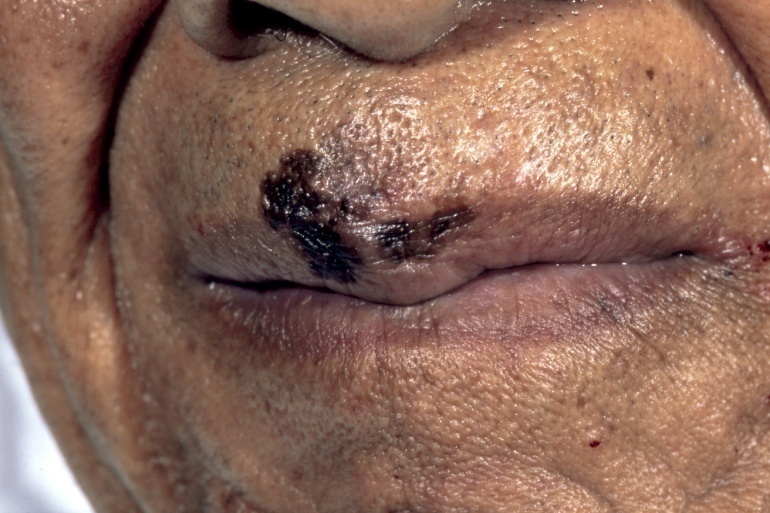
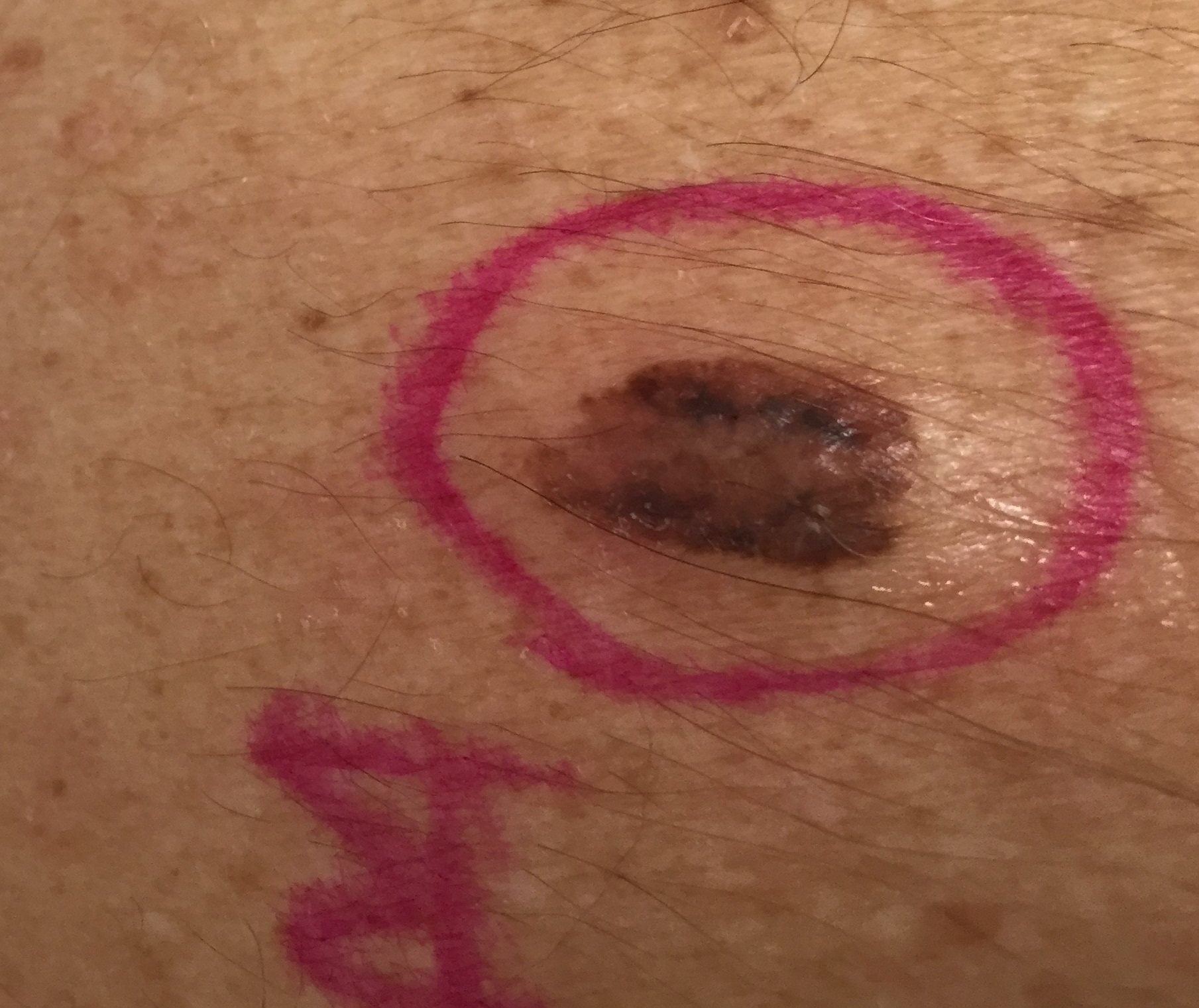
- Melanoma photos: Early stage and late stage melanoma images, and pictures of melanoma on various skin tones.
- Merkel cell carcinoma photos
To catch skin cancer early, you should examine your skin once a month. If you see something NEW, CHANGING OR UNUSUAL – even if it looks nothing like what you see in photos – do not wait! Get it checked by a dermatologist right away. Finding and treating skin cancer early can save your life.
Skin Cancer Image Gallery
What does cancer look like on skin? Below is a selection of photos that give you a general idea about what skin cancers can look like.
- Remember that skin cancers can look quite different from one person to another due to skin tone, size and type of skin cancer and location on the body.
- Skin cancer can be tricky in other ways, too. For example, melanoma is a type of skin cancer that is often pigmented tan, brown, black, even blue. But amelanotic melanoma lacks pigment and appears as a skin-tone or pink lesion.
To sum it up, while photos can be helpful, getting your skin examined by a dermatologist is the most vital step in identifying and treating skin cancer.
Please note: Since not all skin cancers have the same appearance, these photos serve as a general reference for what skin cancer can look like. If you see anything NEW, CHANGING or UNUSUAL on your skin, go get checked by a dermatologist.


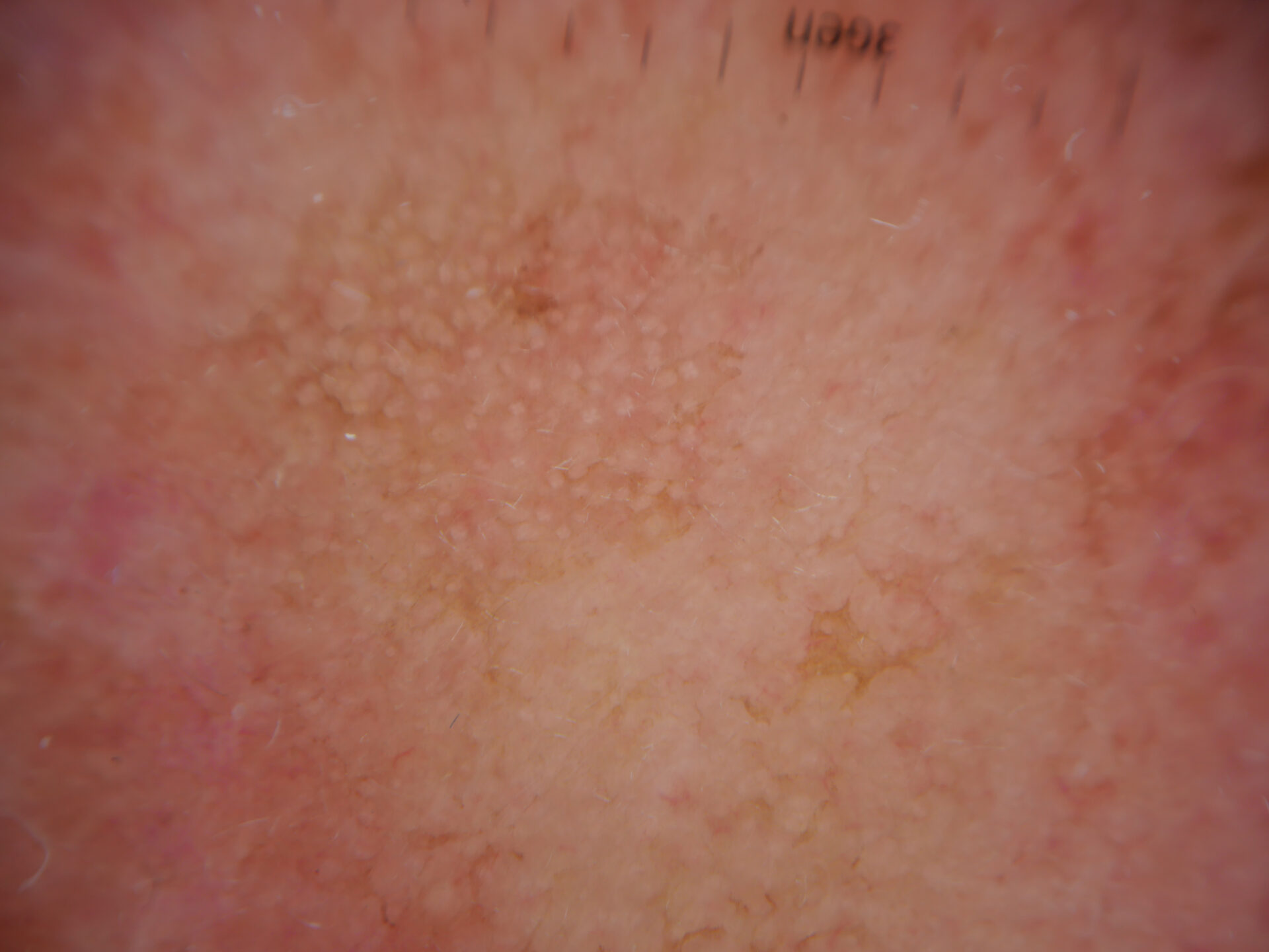
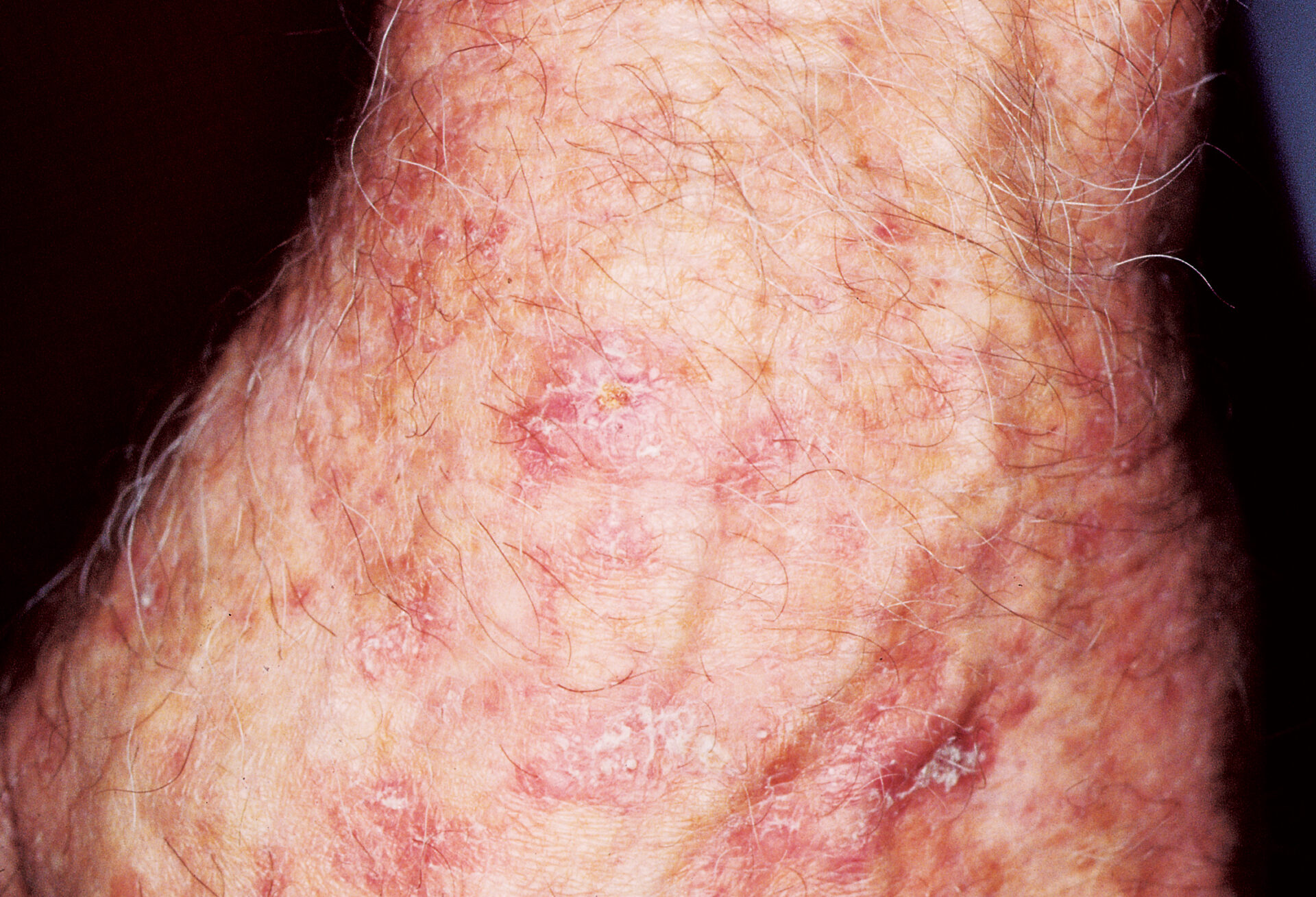
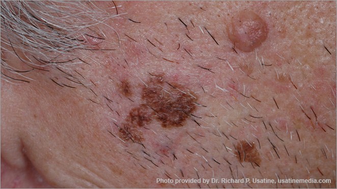
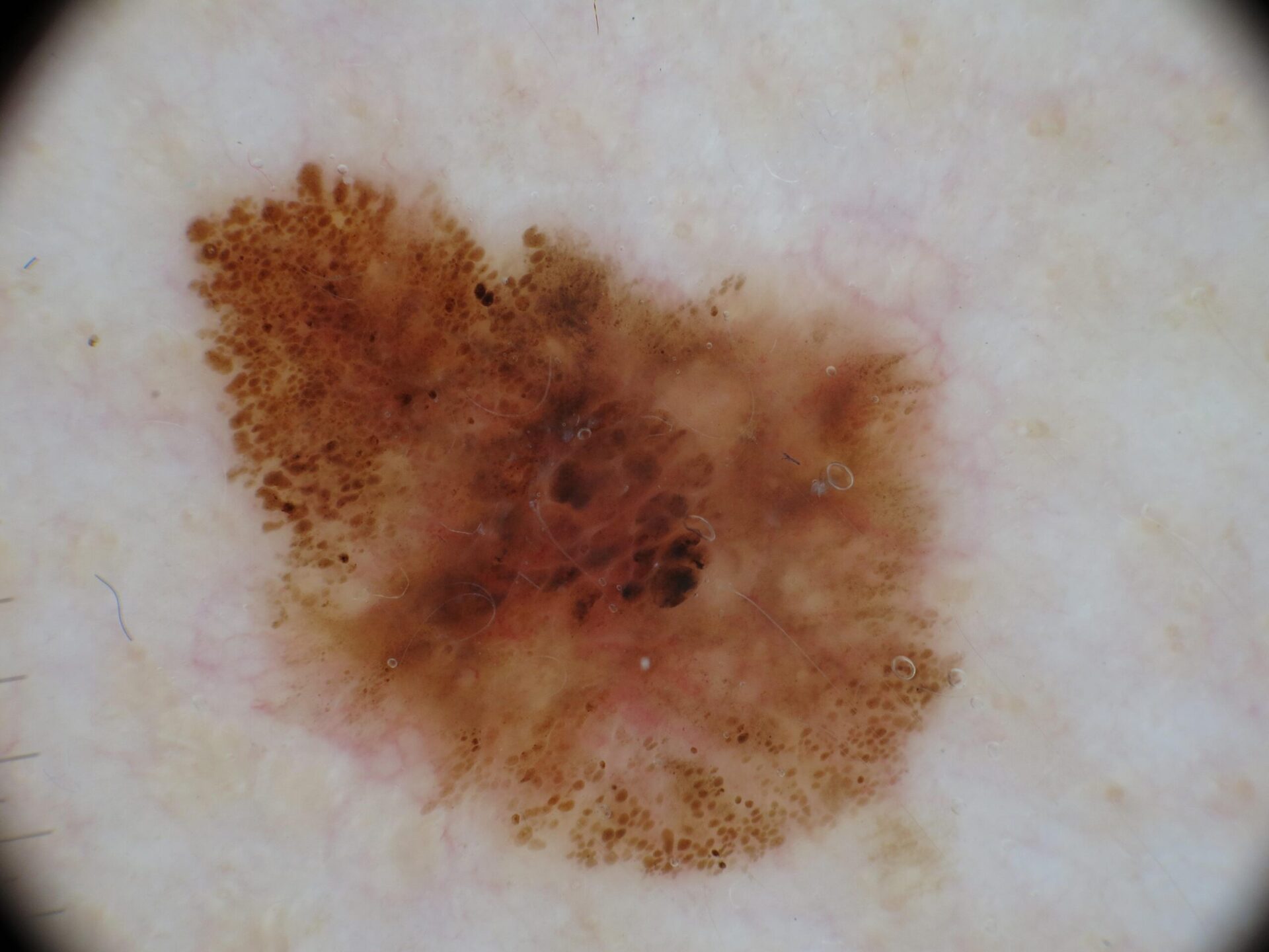
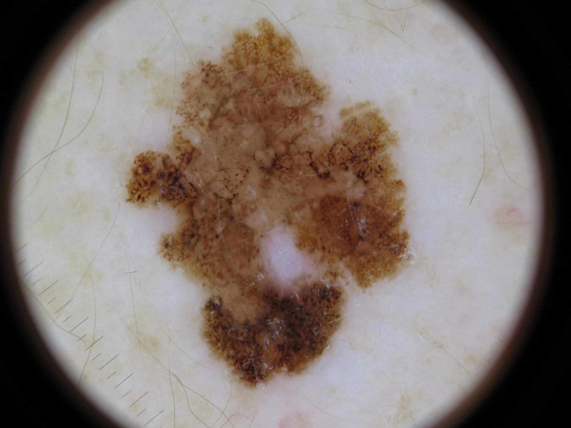
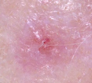
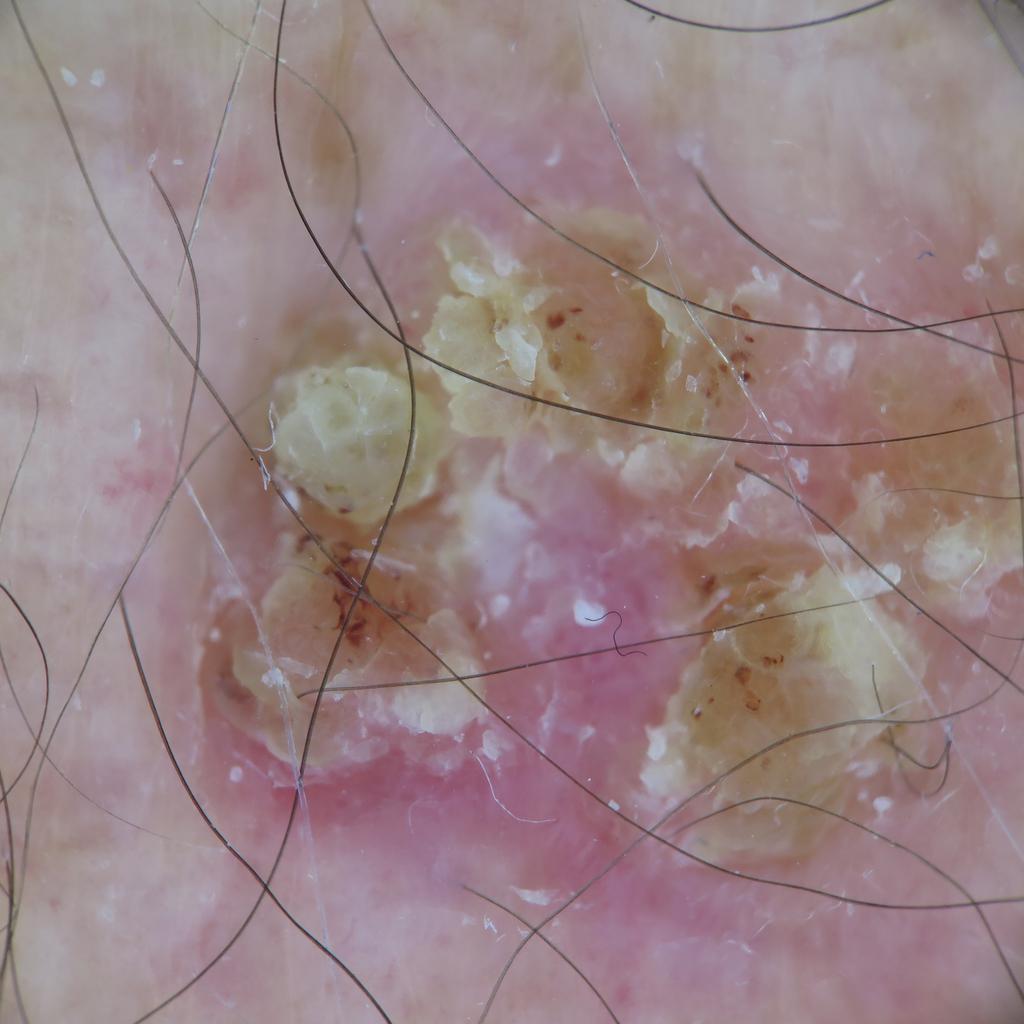
Actinic Keratosis (AK) Photos
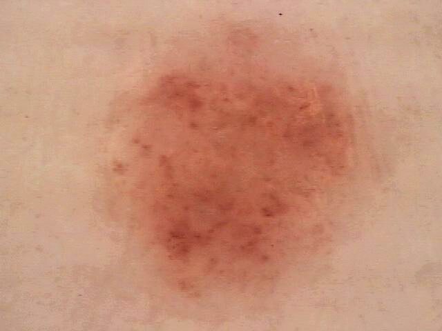
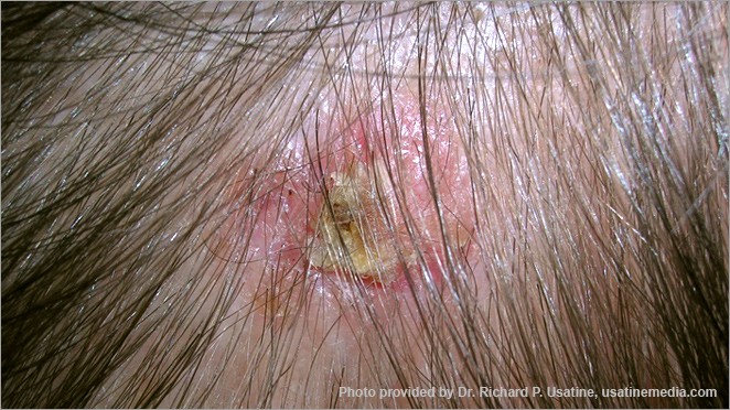
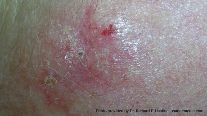
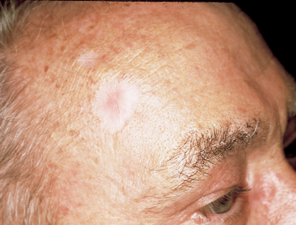
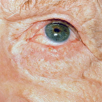
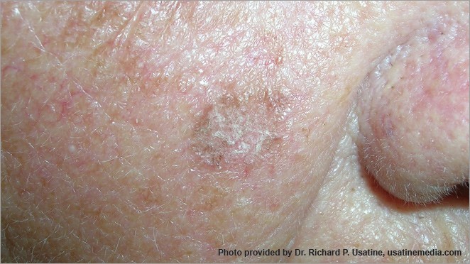
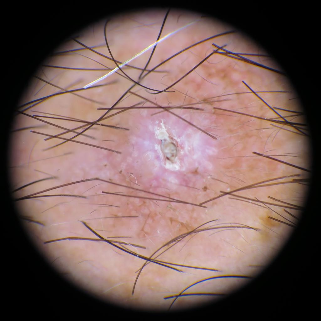
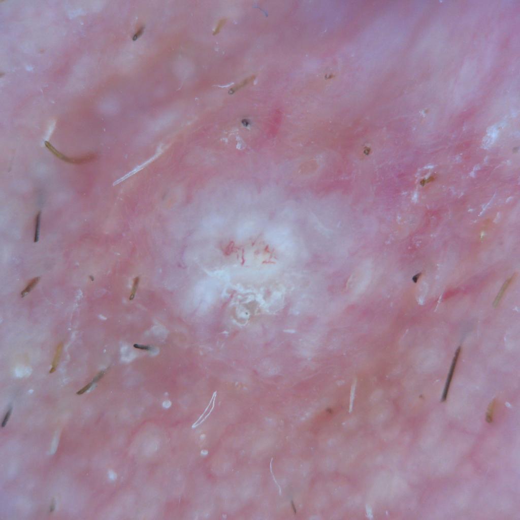
Actinic keratosis on head or neck. Photo: International Skin Imaging Collaboration at isic-archive.com
Actinic keratosis on posterior torso. Photo: International Skin Imaging Collaboration at isic-archive.com
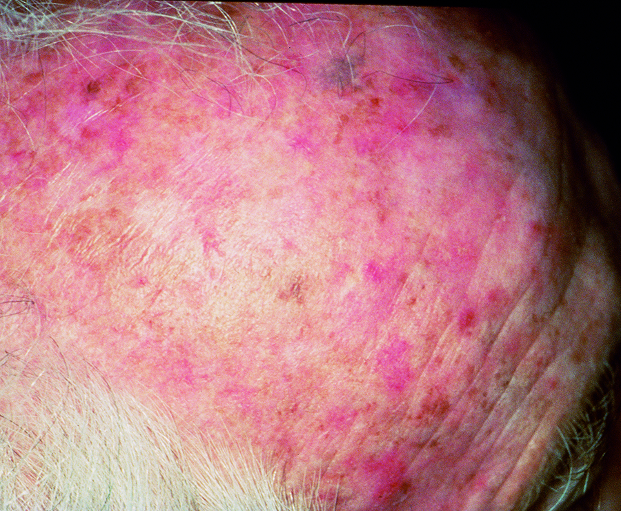


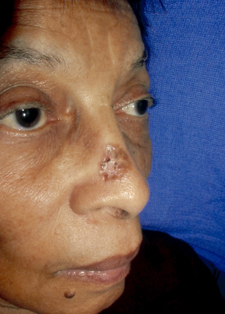
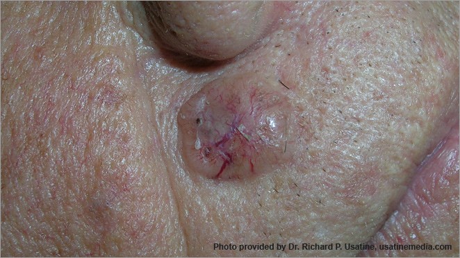
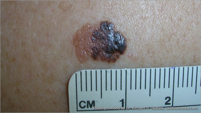
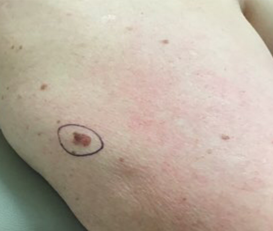
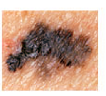
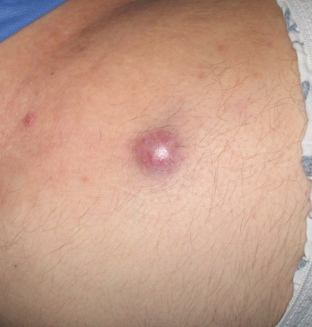
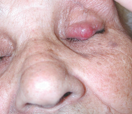
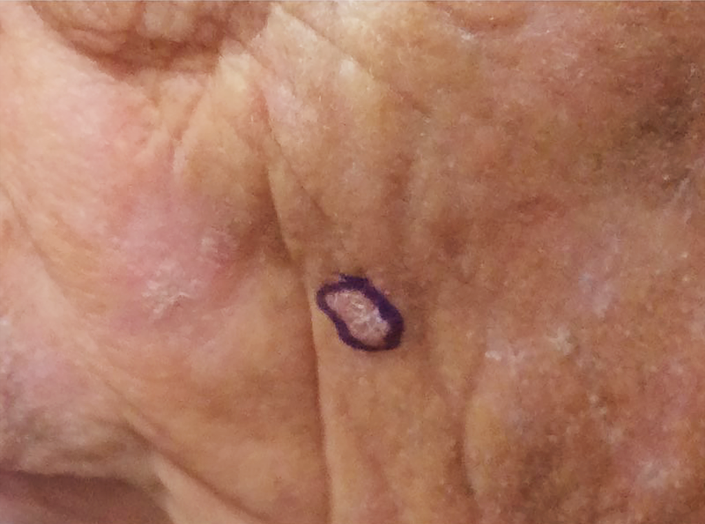
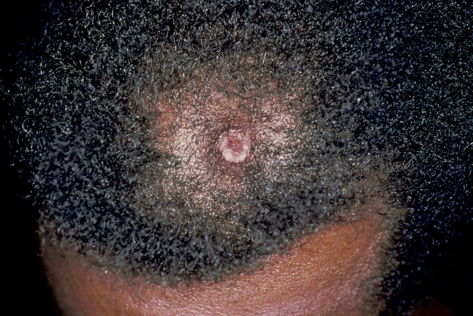
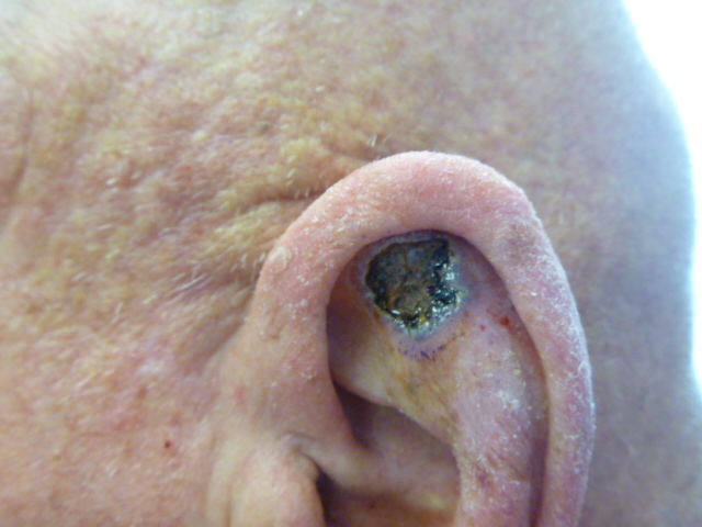
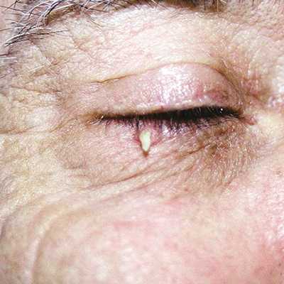
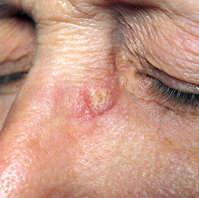
Basal Cell Carcinoma (BCC) Photos
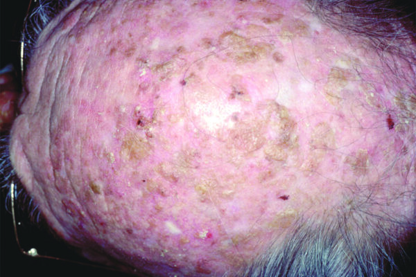
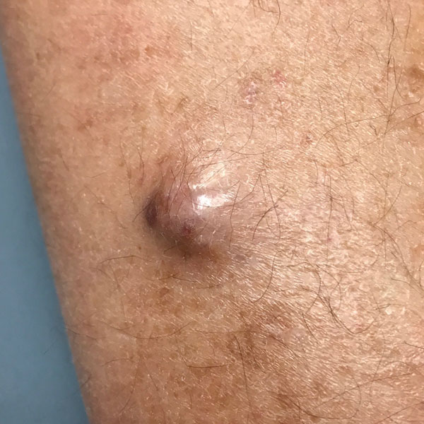
Basal cell carcinoma on the posterior torso. Photo: International Skin Imaging Collaboration at isic-archive.com
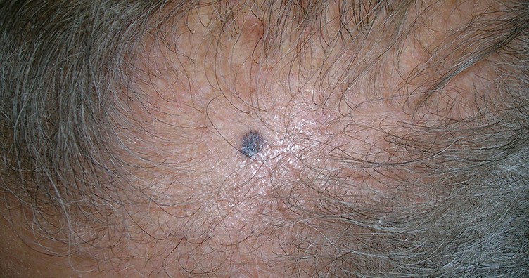
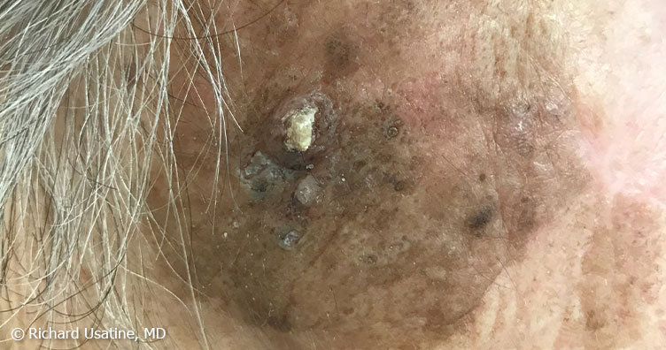
Basal cell carcinoma on the neck. Photo: International Skin Imaging Collaboration at isic-archive.com
Basal cell carcinoma on the anterior torso. Photo: International Skin Imaging Collaboration at isic-archive.com
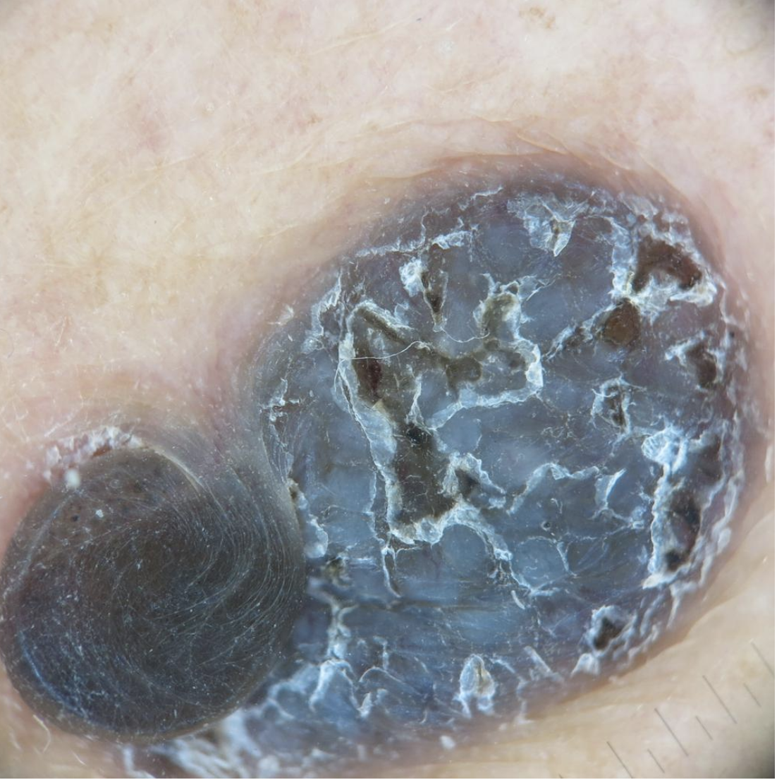
Basal cell carcinoma on the anterior torso. Photo: International Skin Imaging Collaboration at isic-archive.com
Basal cell carcinoma on the anterior torso. Photo: International Skin Imaging Collaboration at isic-archive.com
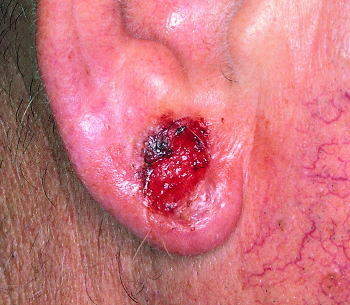
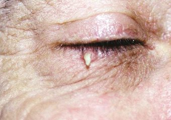
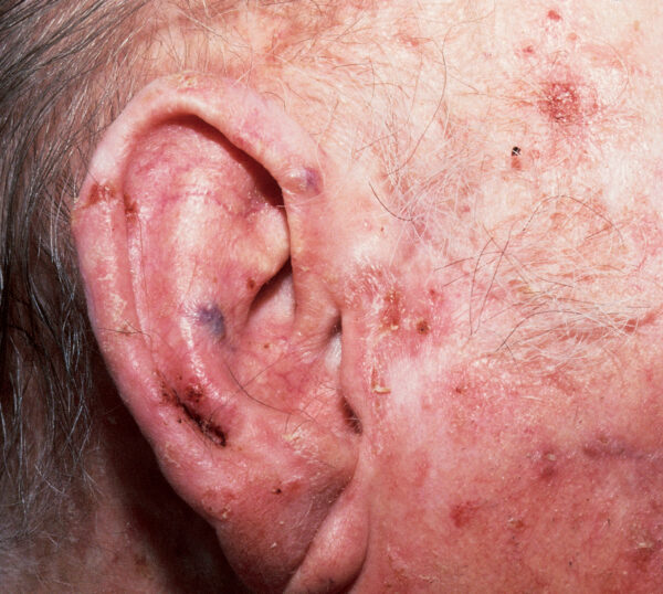
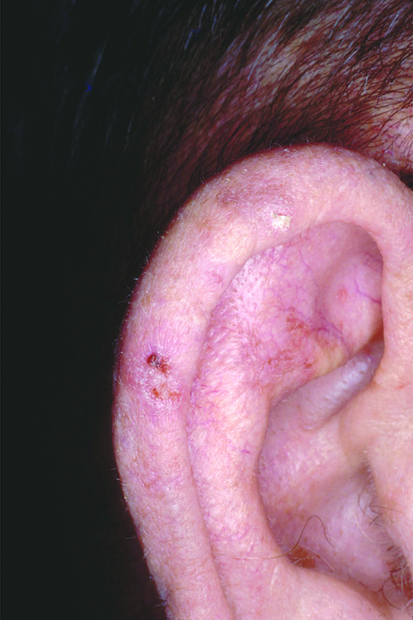
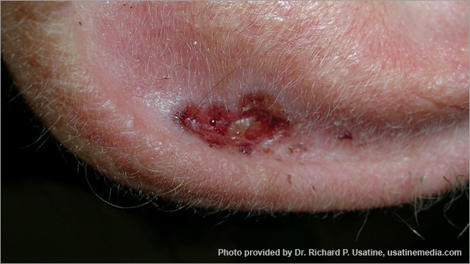
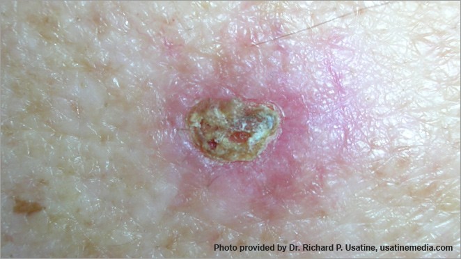
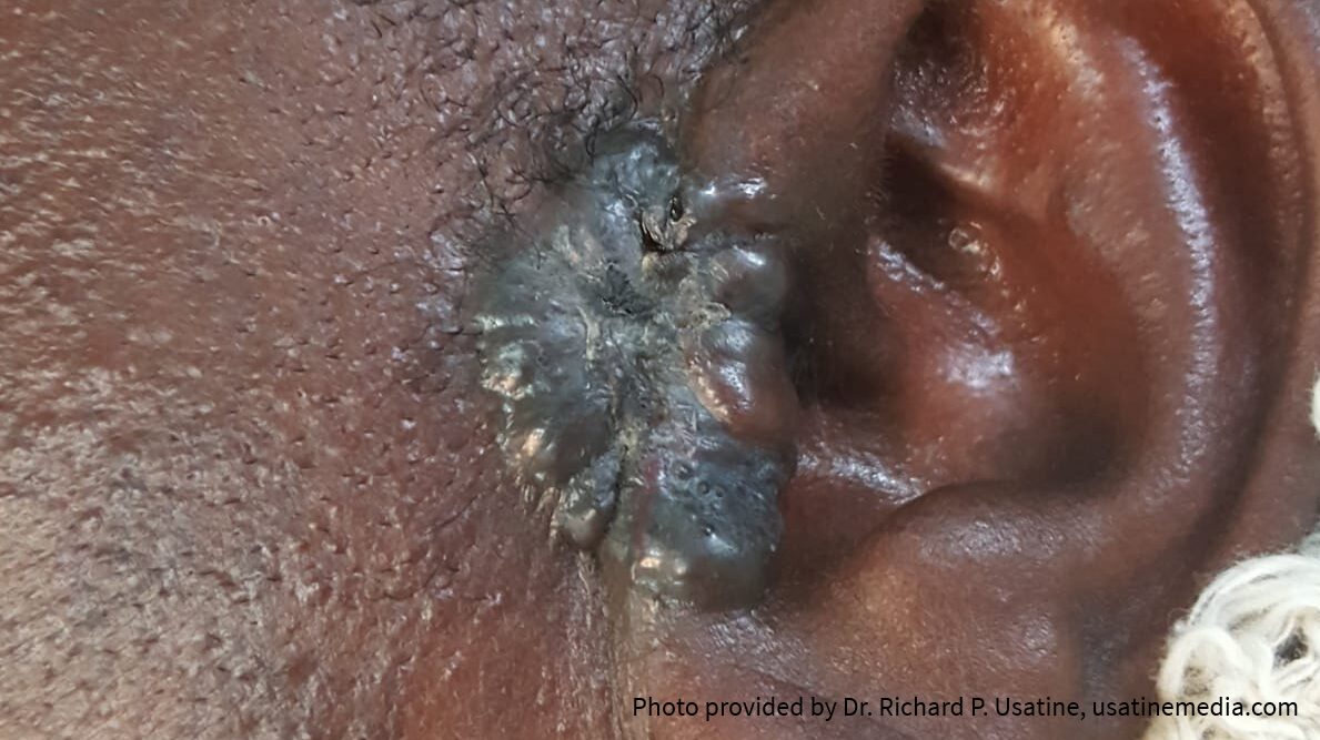
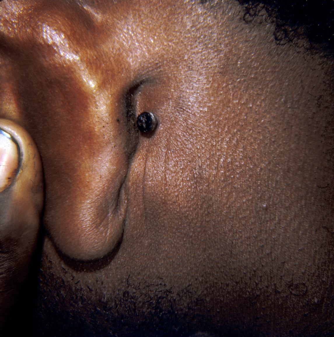
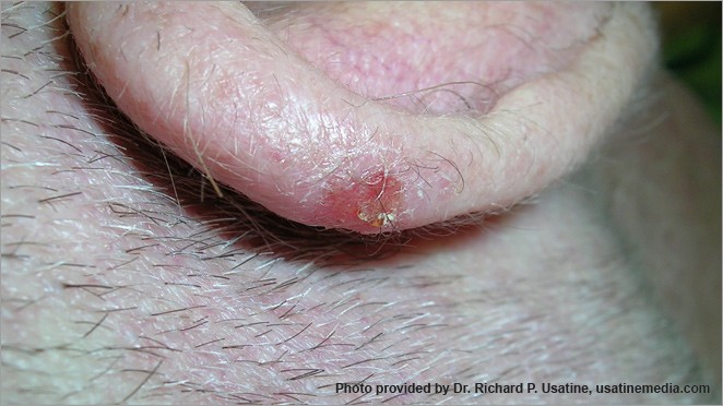
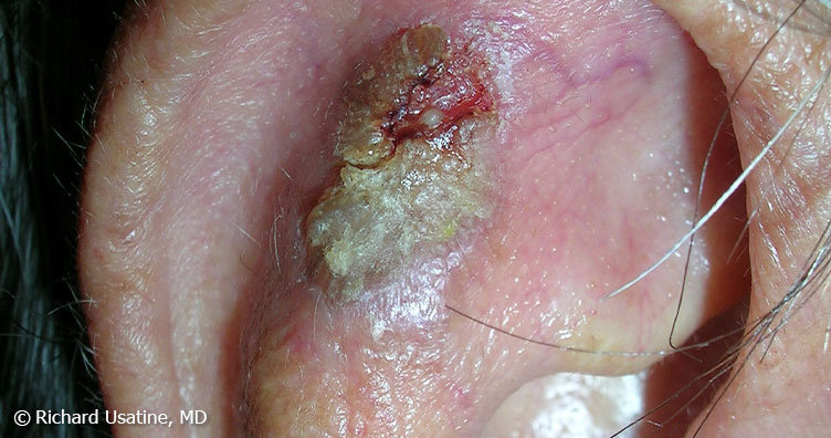
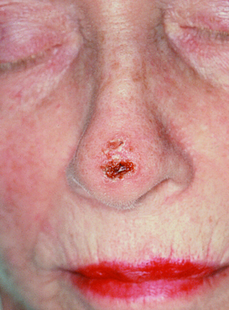
Basal cell carcinoma presenting as an open sore on the ear. Photo: International Skin Imaging Collaboration at isic-archive.com
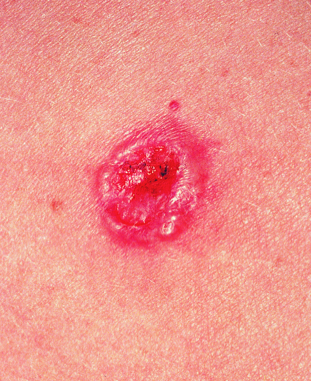
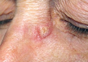
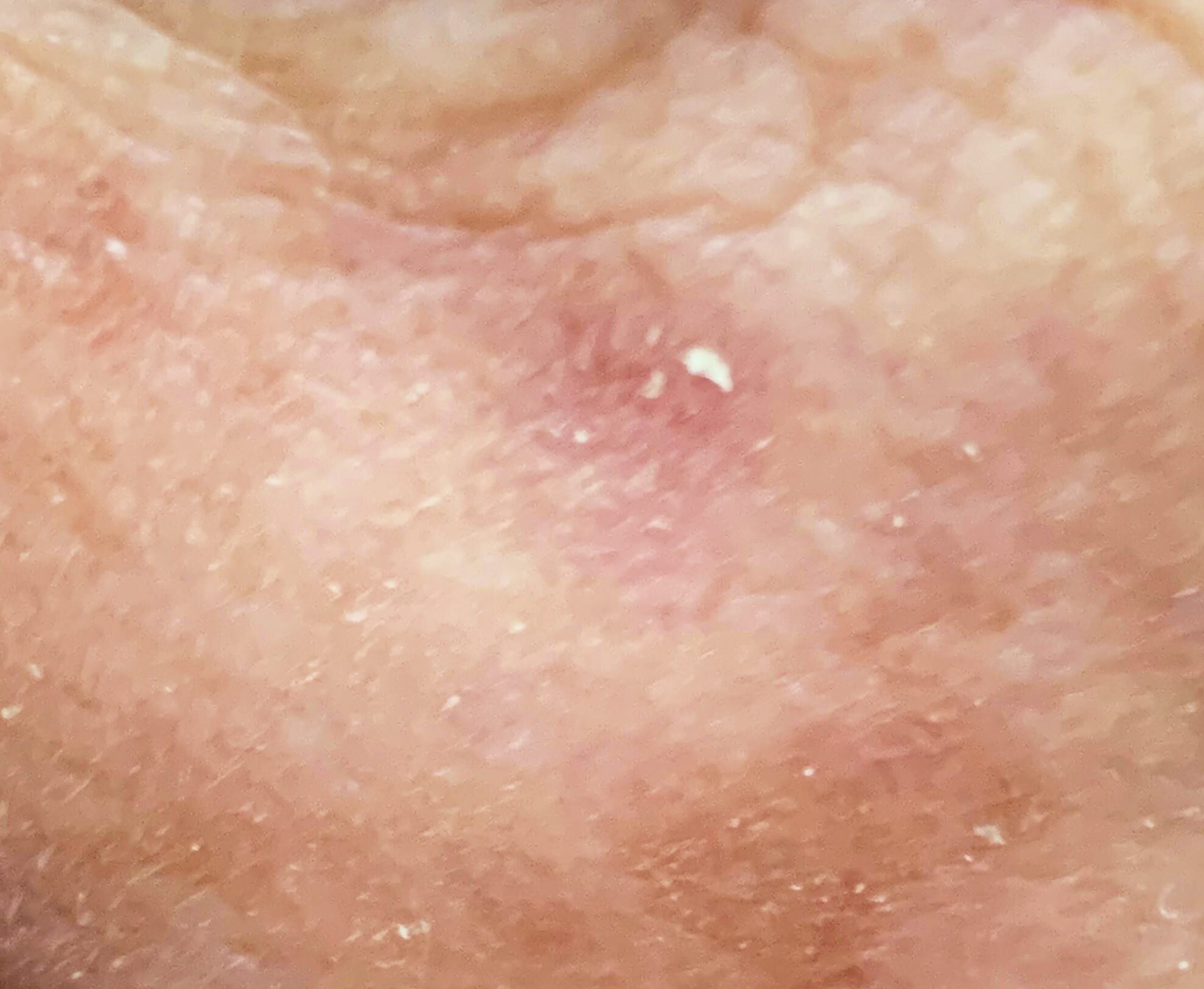
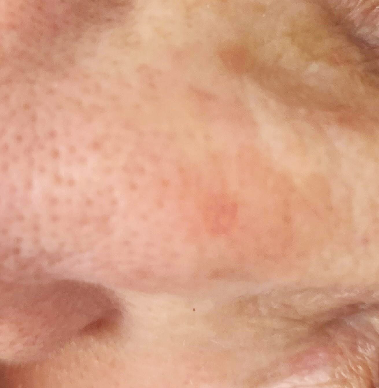
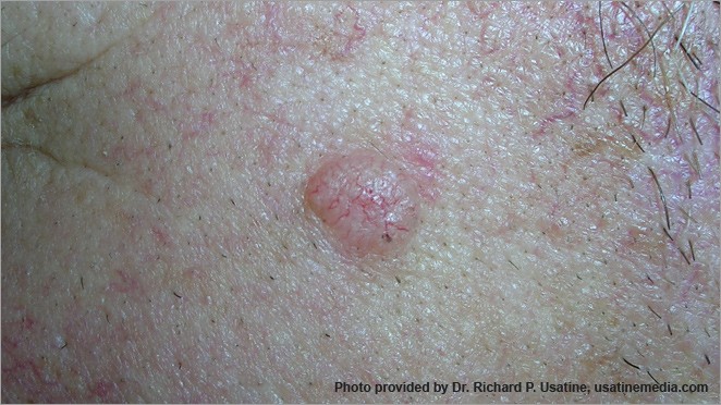
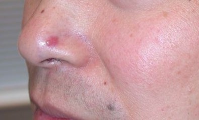
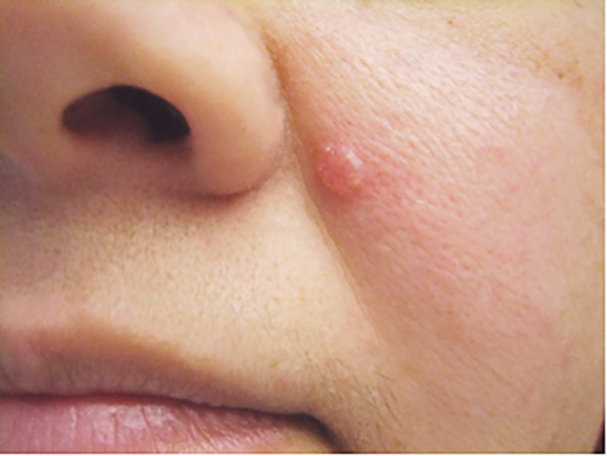
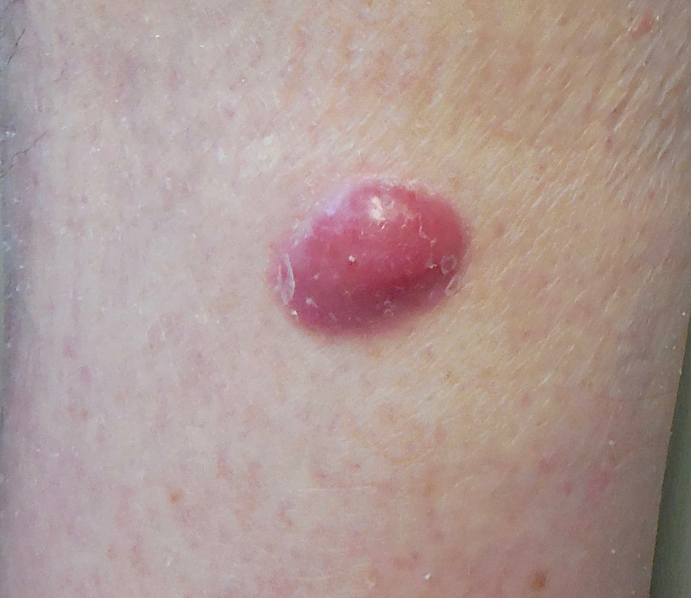
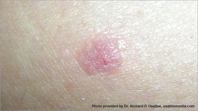
Basal cell carcinoma presenting as a pink growth. Photo: International Skin Imaging Collaboration at isic-archive.com
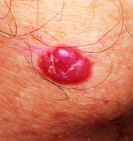
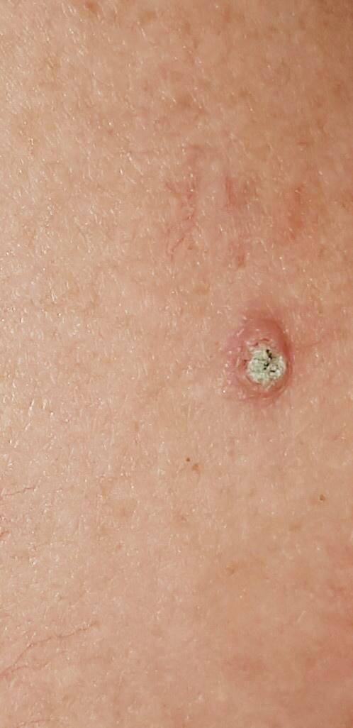
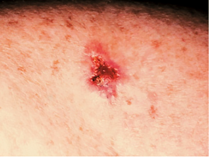
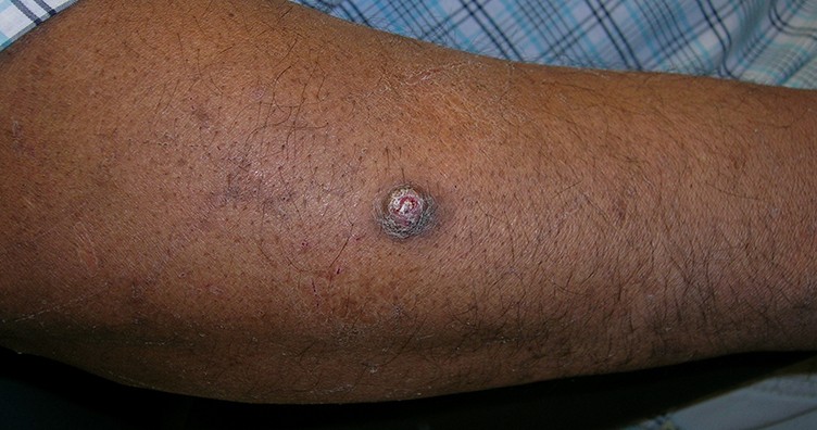
Basal cell carcinoma on the leg. Photo: International Skin Imaging Collaboration at isic-archive.com
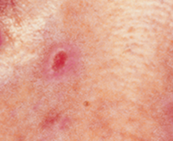
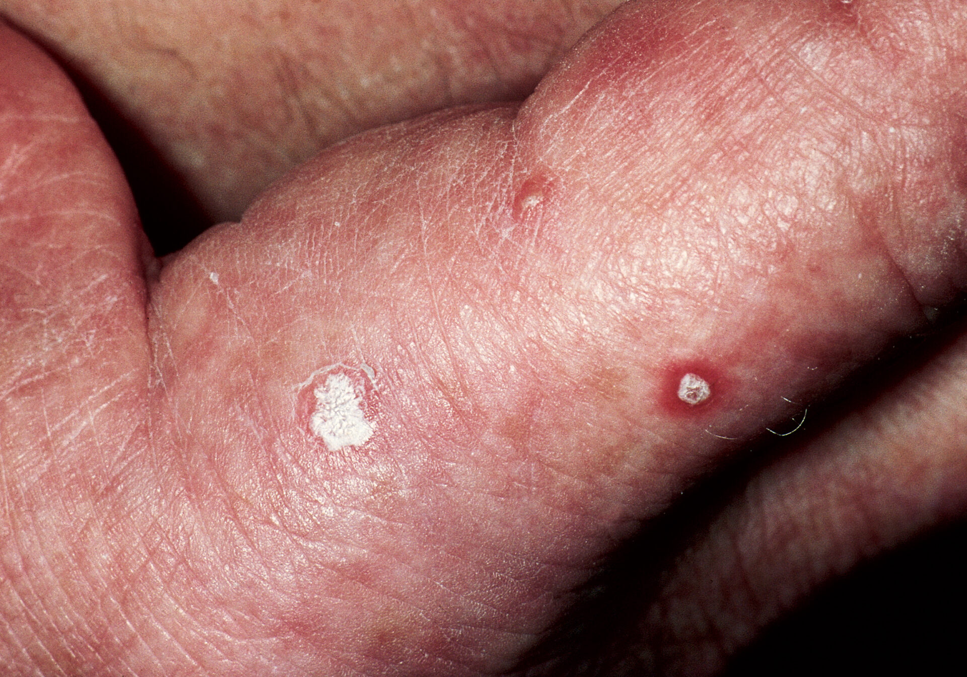
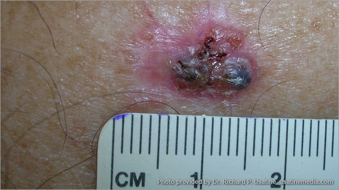
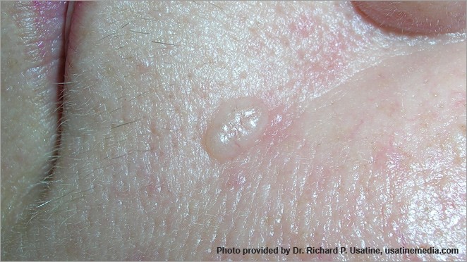
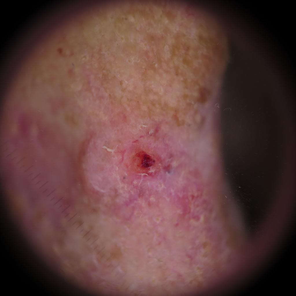
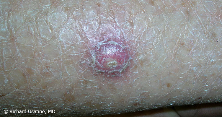
A small pink growth with a slightly raised, rolled edge and a crusted indentation in the center. (BCC)
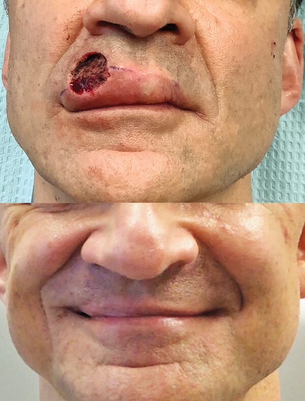
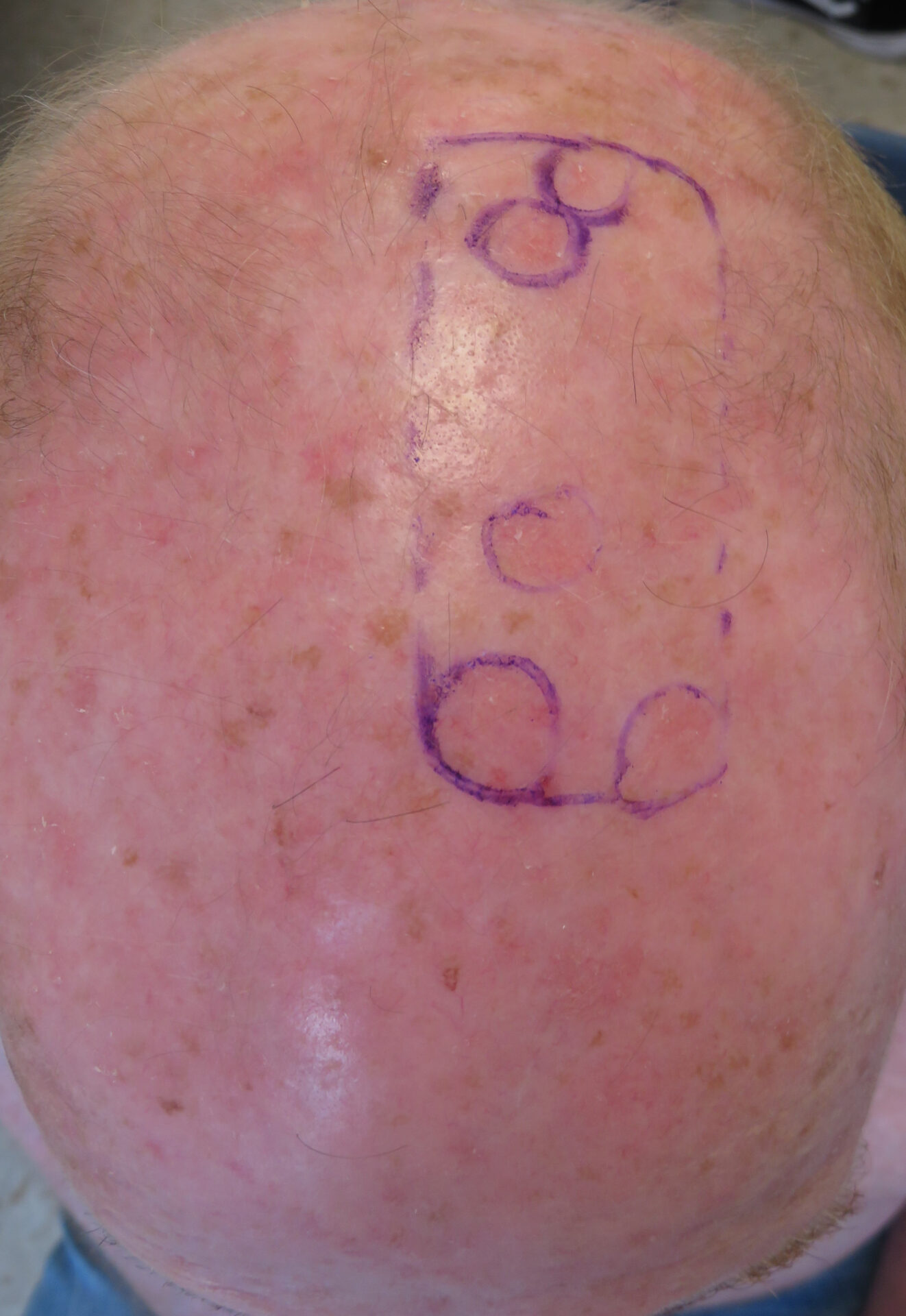
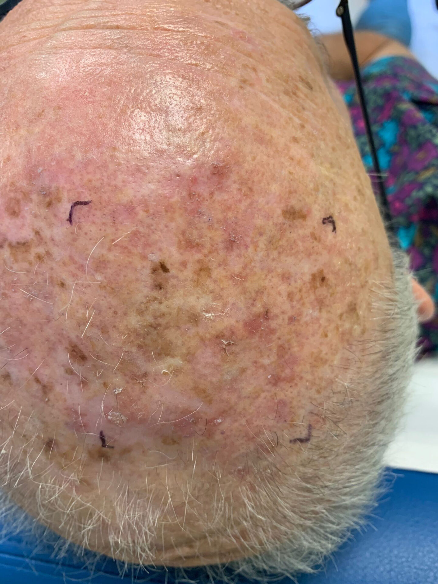
Basal Cell Carcinoma (BCC) Case Study
Inconvenient BCC, Good Outcome
Deborah S. Sarnoff, MD
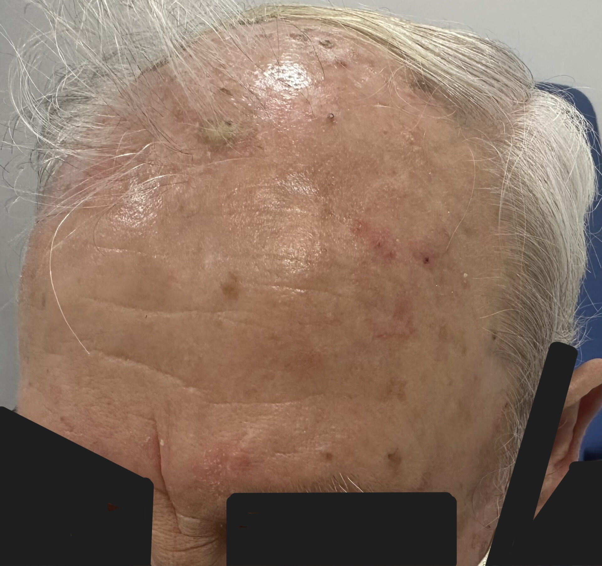
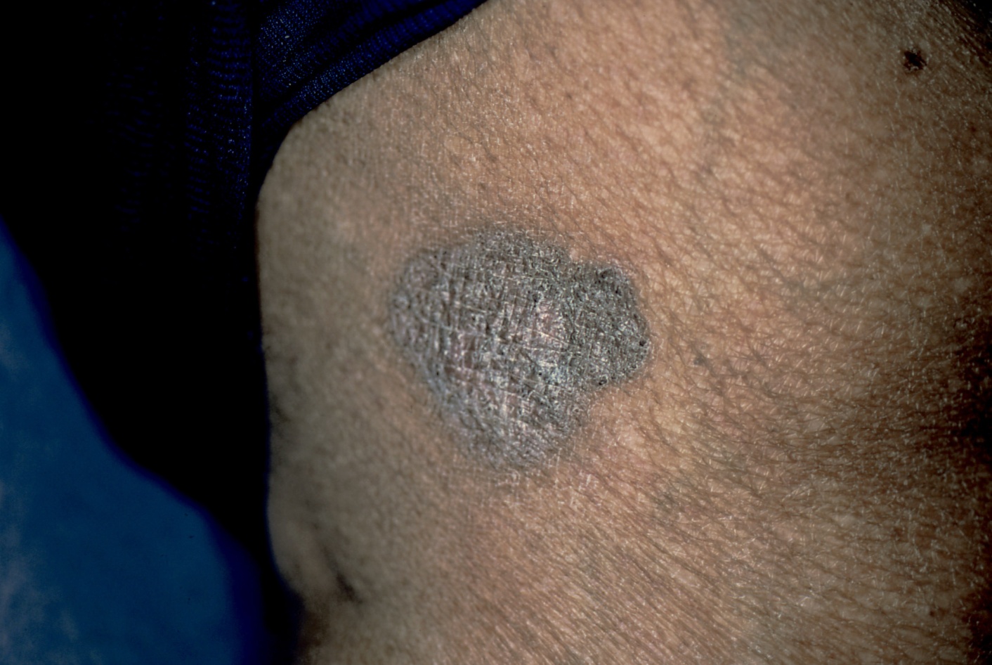
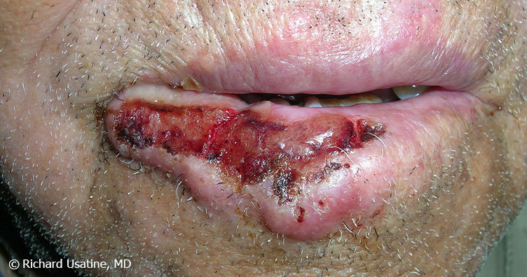
A patient in his 40s with the genetic condition known as Gorlin syndrome had been treated for many basal cell carcinomas. During the COVID pandemic, he noticed a new bump on his upper lip. It didn’t seem serious to him, so, like many patients during that time, he did not see his dermatologist. When he finally did get checked, it was indeed a BCC and had grown deep enough to require several stages of Mohs surgery. The surgery eliminated the cancer but left the lower part of his face disfigured (top photo). Dr. Sarnoff’s business partner and husband, plastic surgeon Robert H. Gotkin, MD, was able to reconstruct the area with minimal scarring and an excellent cosmetic result (bottom photo, after healing). Read the full story here: http://www2.skincancer.org/blog/we-dont-want-to-scare-you-but/
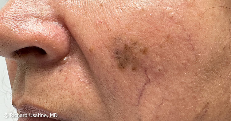
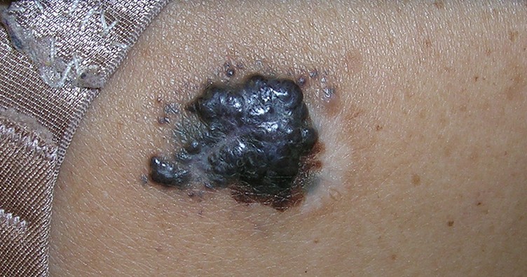
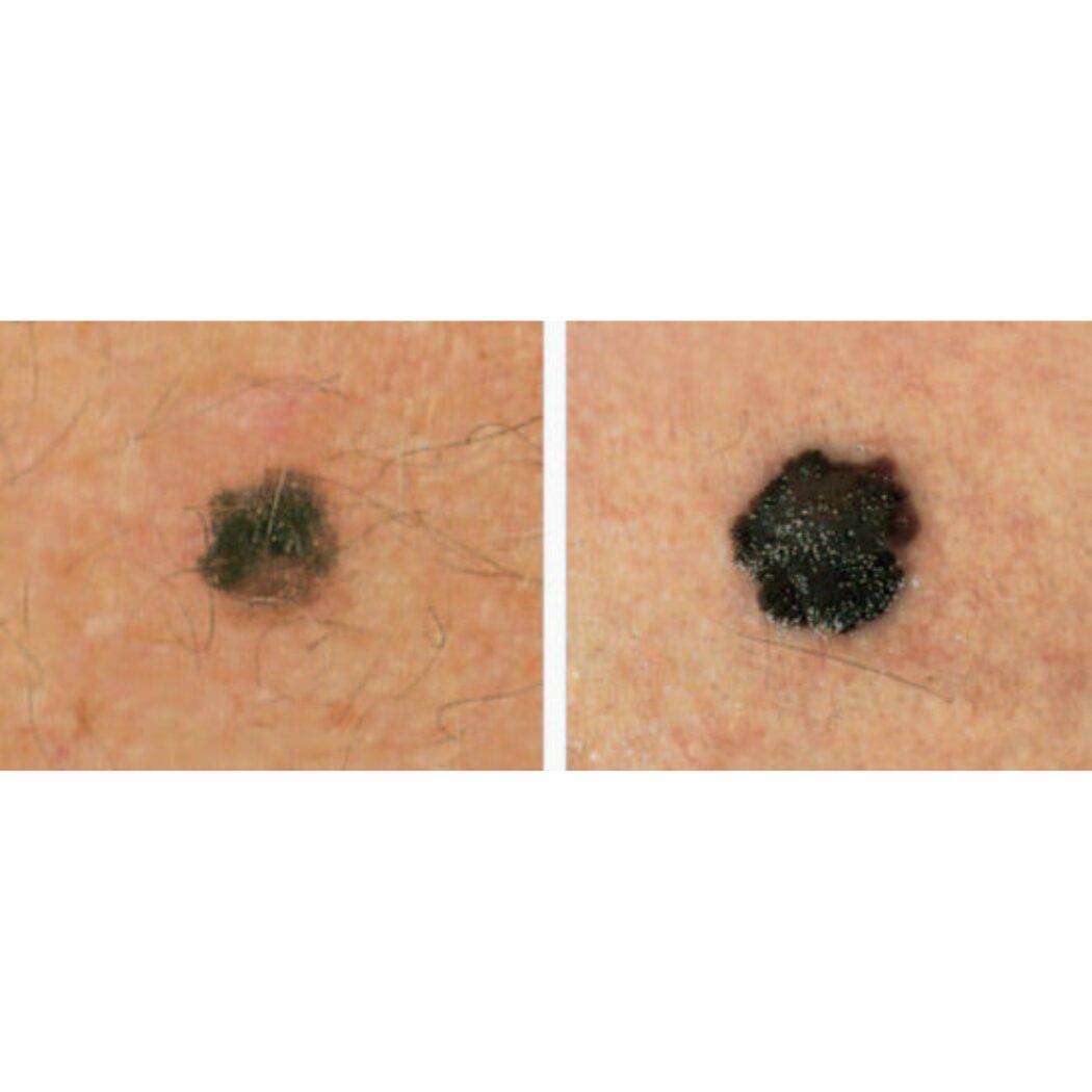
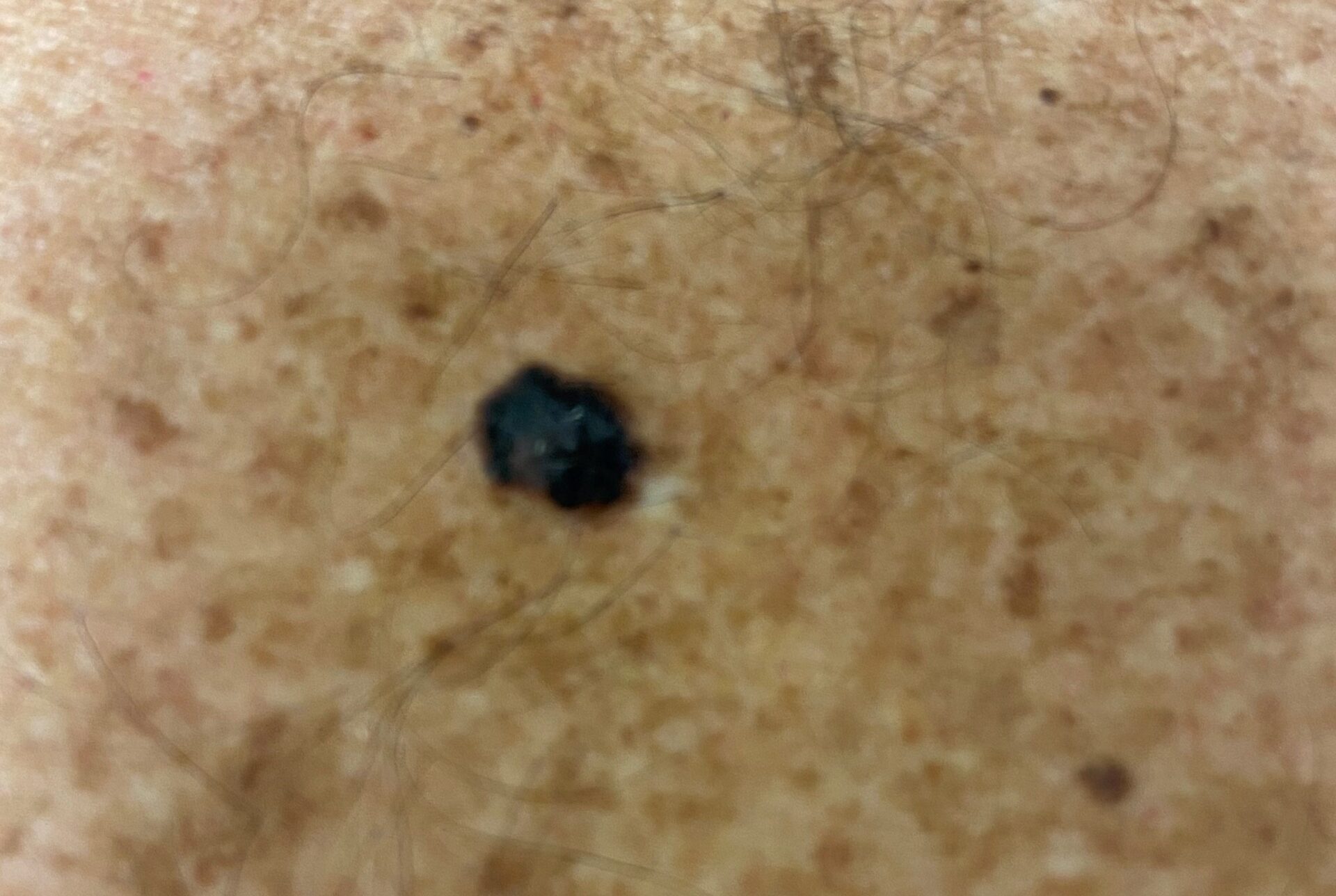
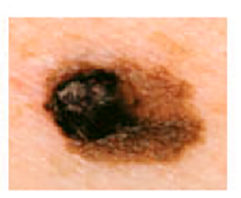
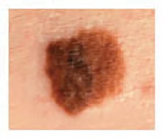
Melanoma Photos

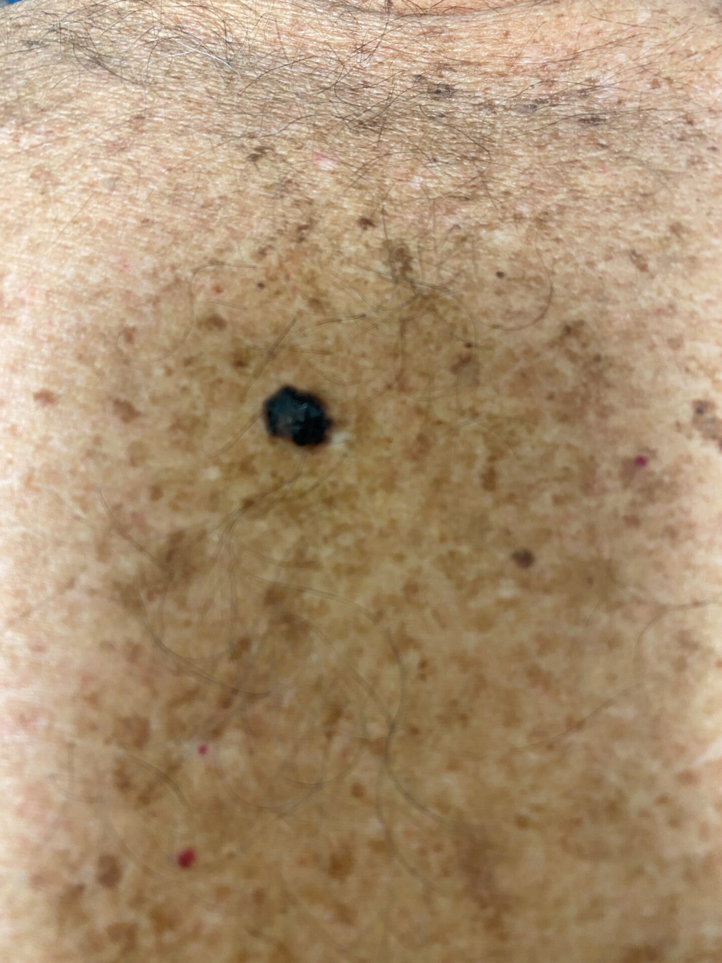
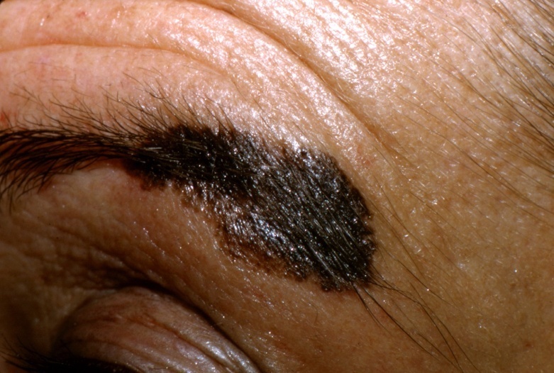
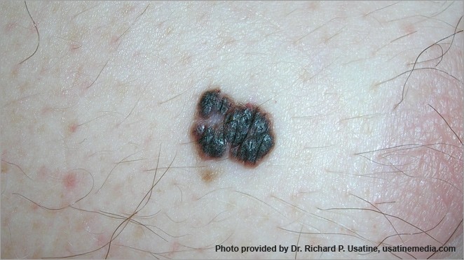
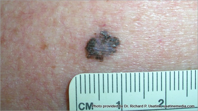
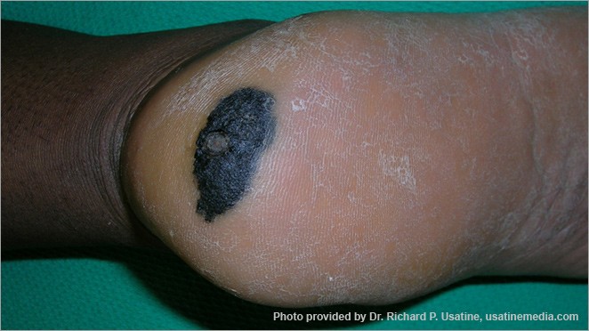
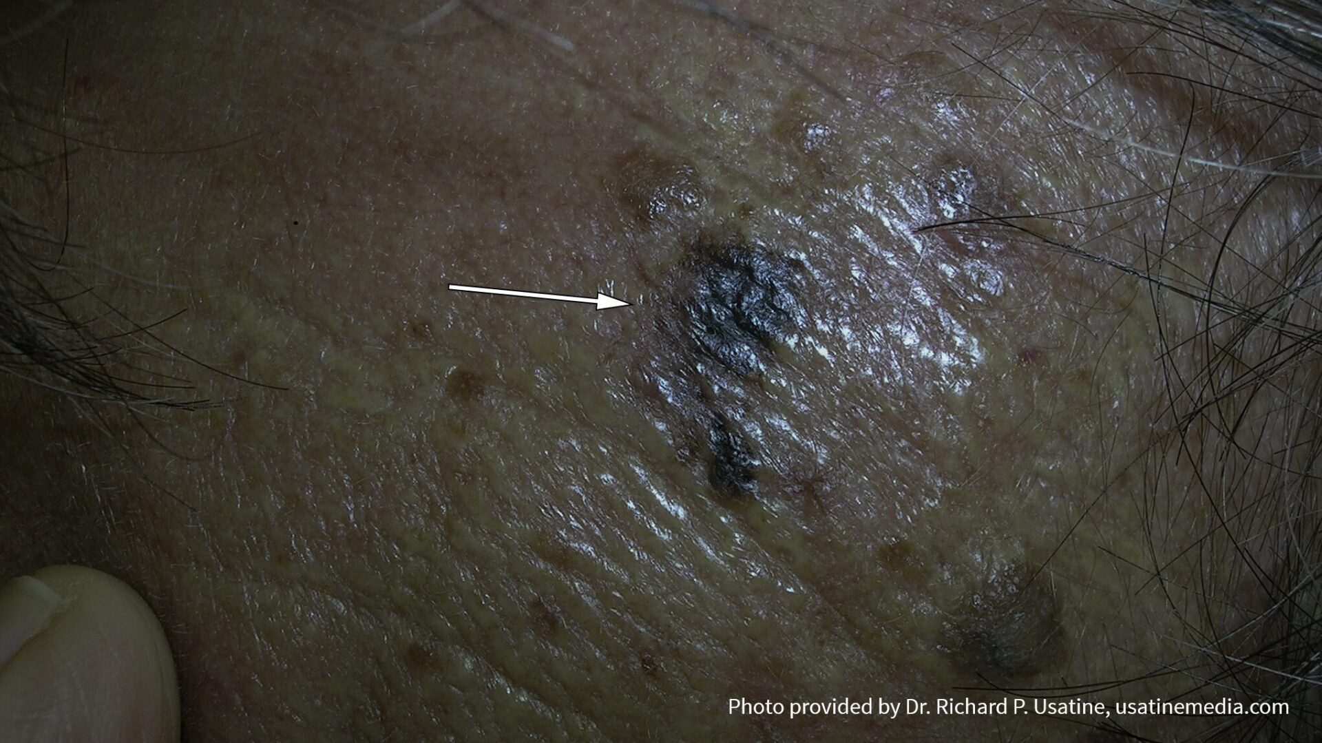
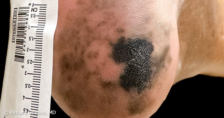
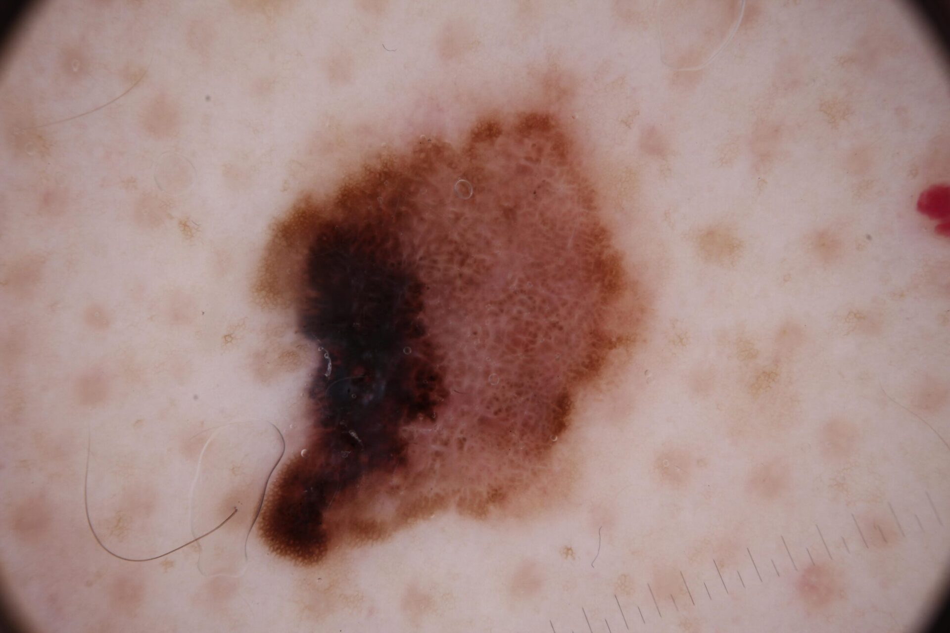
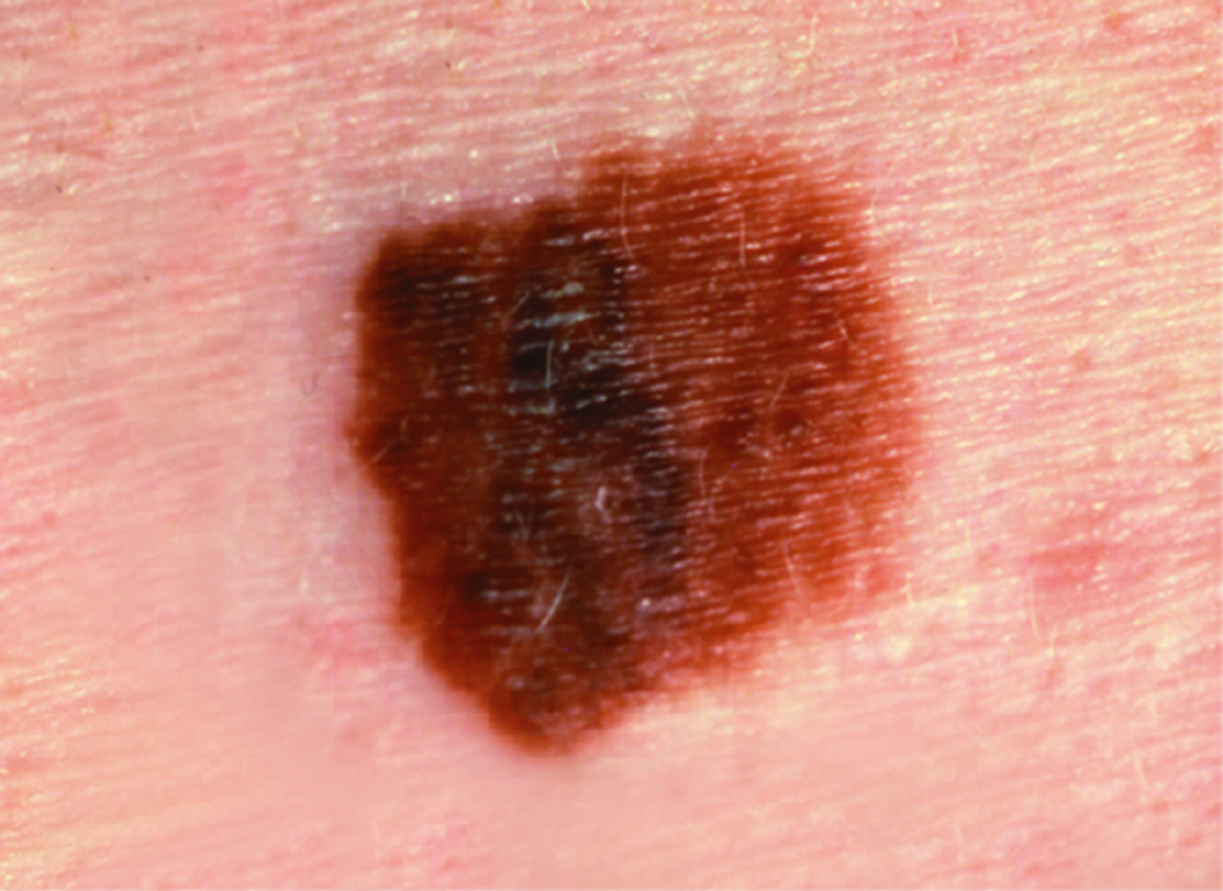
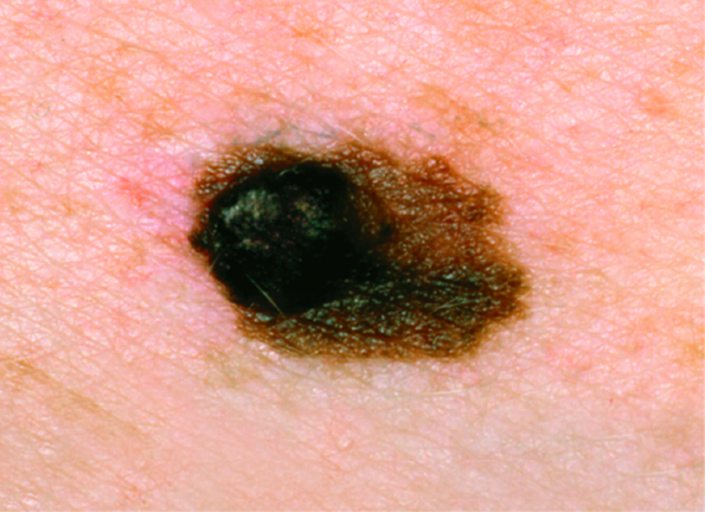
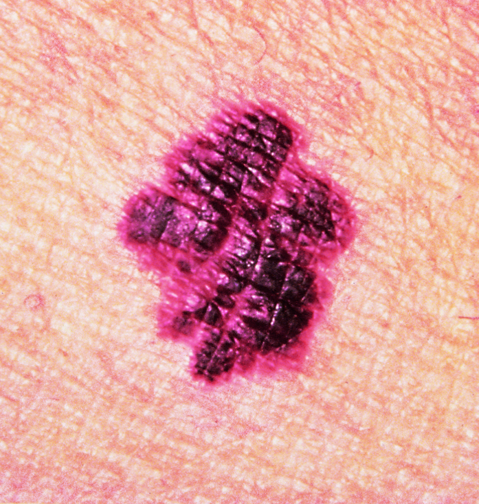
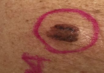
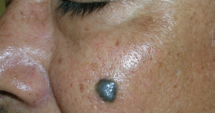
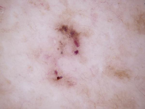
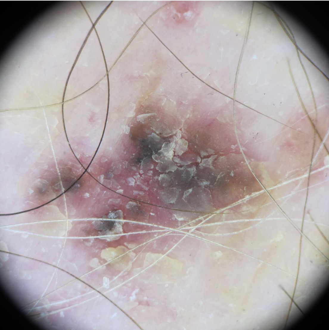
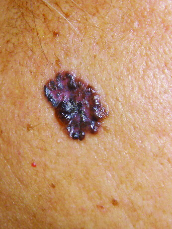
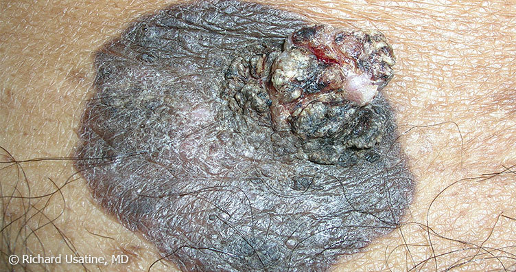
Nodular melanoma on the leg of a Native American woman.
Photo: International Skin Imaging Collaboration
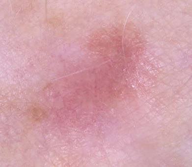
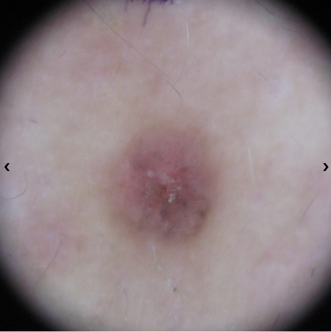
Amelanotic melanomas may be pinkish-looking, reddish, purple, normal skin color or essentially clear and colorless.

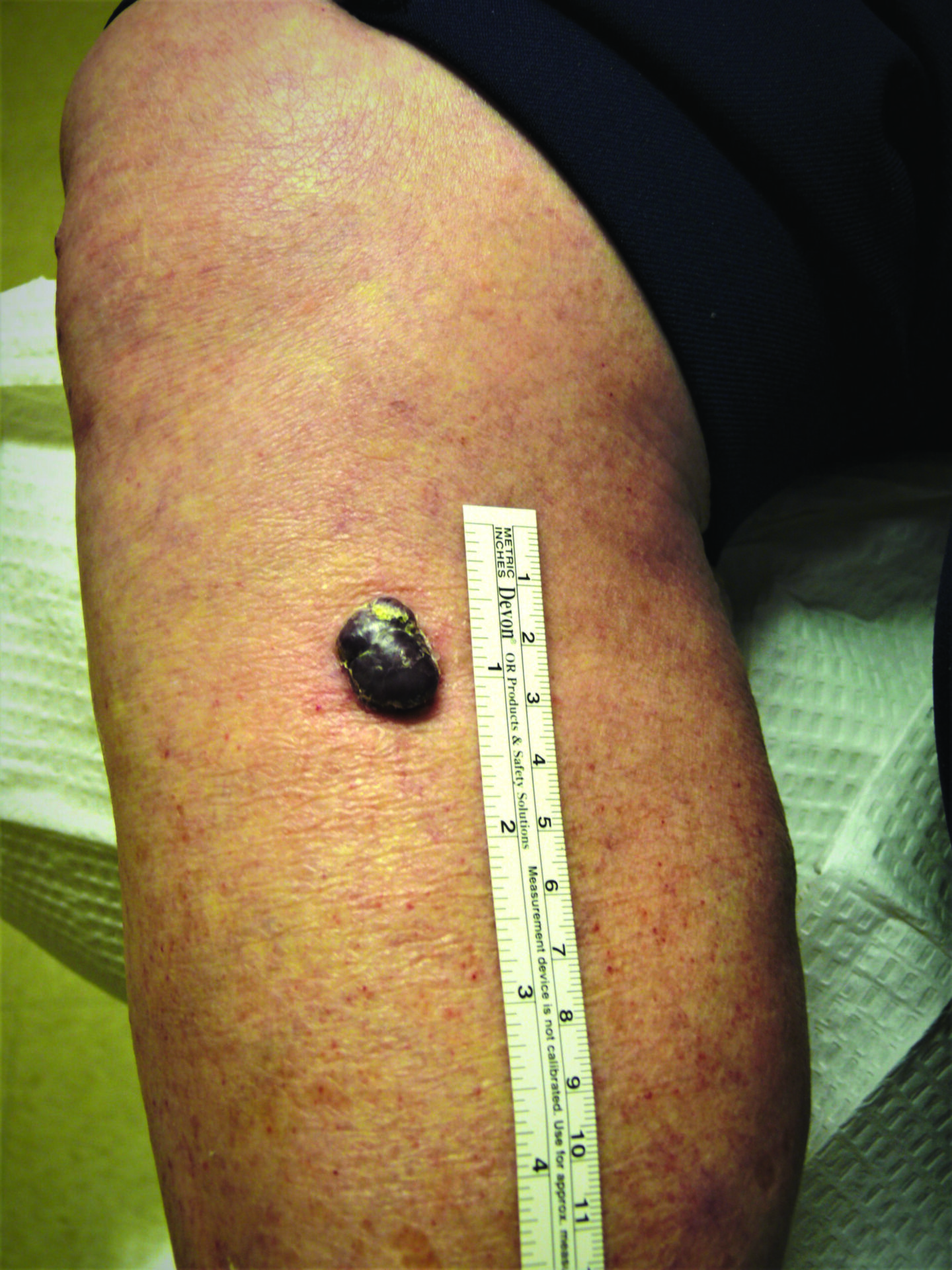
Melanoma Case Study
Not a “Boil” but an Advanced Melanoma
Deborah S. Sarnoff, MD
The power of denial can be strong. This man believed the large, dark growth on his back was a boil, despite oozing and bleeding for months. He kept bandaging it and thought it was just slow to heal. When he was finally referred to Dr. Sarnoff, a biopsy and other tests revealed a melanoma so large and deep that it had already spread to the man’s liver and brain. He was referred to an oncologist and began an immunotherapy regimen that did not appear to be working, which happens with some patients. (Thanks to recent innovations, there may be other treatment options, including participation in a clinical trial.) This is a powerful reminder of the importance of early detection.
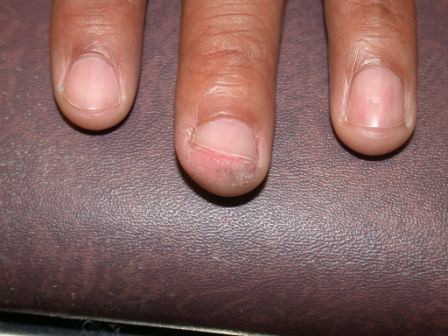
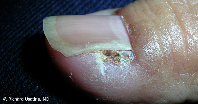
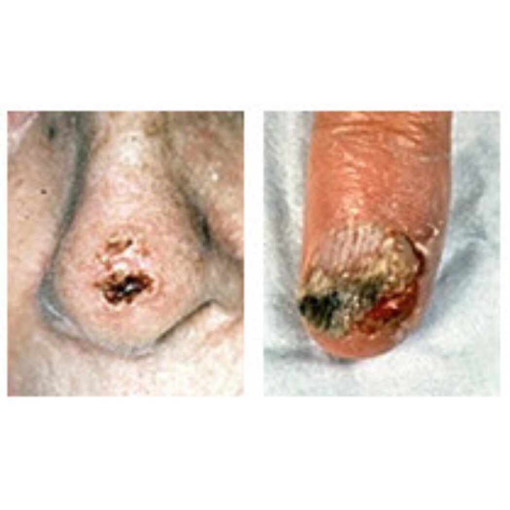
Merkel Cell Carcinoma (MCC) Pictures

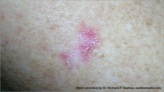
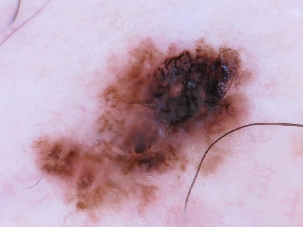
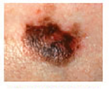
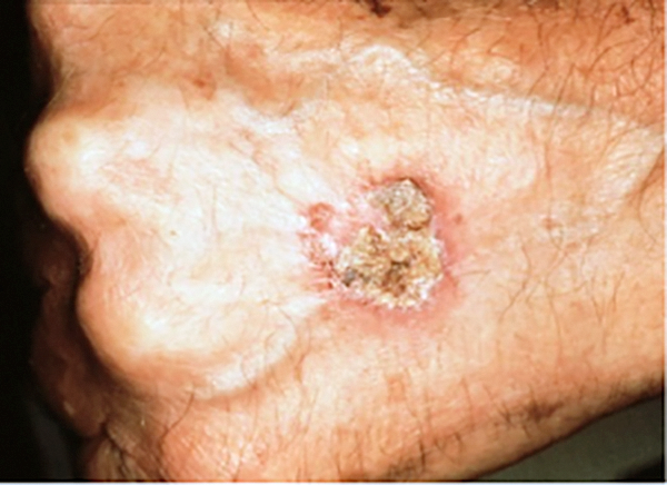
Squamous Cell Carcinoma (SCC) Photos
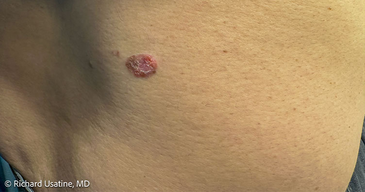
Squamous cell carcinoma on forearm of man with darker skin tone
Photo credit: Richard P. Usatine, MD.
Squamous cell carcinoma on the nose. Photo: International Skin Imaging Collaboration at isic-archive.com
Squamous cell carcinoma on the head. Photo: International Skin Imaging Collaboration at isic-archive.com
Squamous cell carcinoma on the neck. Photo: International Skin Imaging Collaboration at isic-archive.com
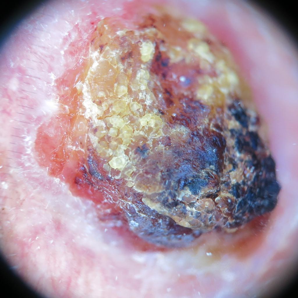
Squamous cell carcinoma on the head. Photo: International Skin Imaging Collaboration at isic-archive.com
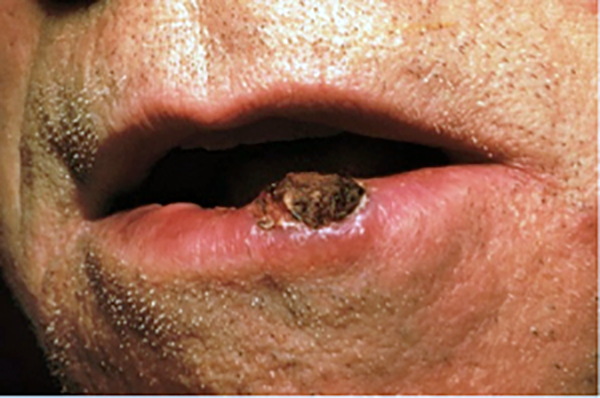
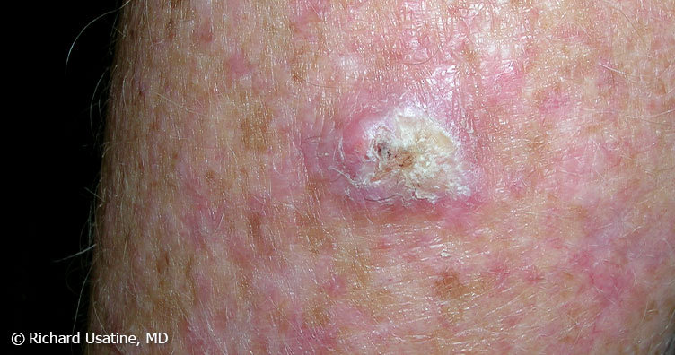
An elevated growth with a central depression that occasionally bleeds. It may rapidly increase in size. (SCC)

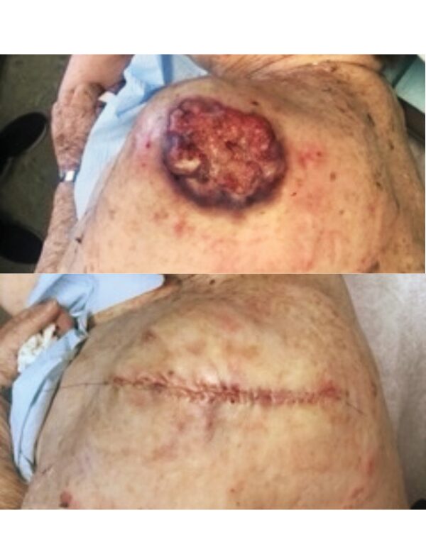
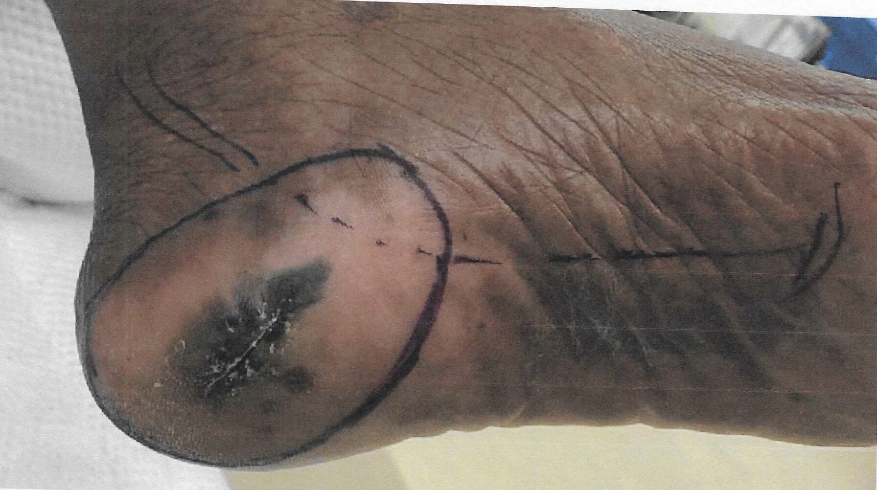
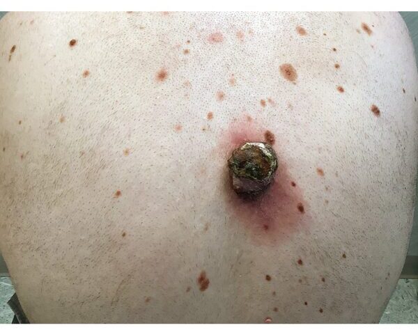
Squamous Cell Carcinoma (SCC) Case Study
A Large Squamous Cell Carcinoma
Deborah S. Sarnoff, MD
Before being admitted to an assisted living facility, an elderly widow was required to have a thorough physical. The doctor (and her adult children who had not seen her undressed in years), were shocked to see a large, raised open wound on her left shoulder and back. For years, she had been covering it and hiding it under clothes. She didn’t want to rock the boat or make a fuss. The lesion (top photo) turned out to be a large SCC, the second most common type of skin cancer. The tumor required extensive surgery followed by radiation. The patient was extremely lucky: Her cancer was eliminated, and Dr. Sarnoff’s business partner and husband Robert H. Gotkin, MD, expertly closed the wound (bottom photo). Dr. Sarnoff strongly advocates robust treatment for elderly people with skin cancer. The earlier it is diagnosed, the less onerous the treatment, and it can make a huge difference in a person’s quality of life.
Rare Skin Cancers
Please visit our Rare skin cancers page for more information and pictures of rare skin cancers such as:
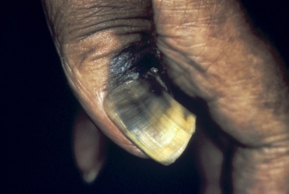
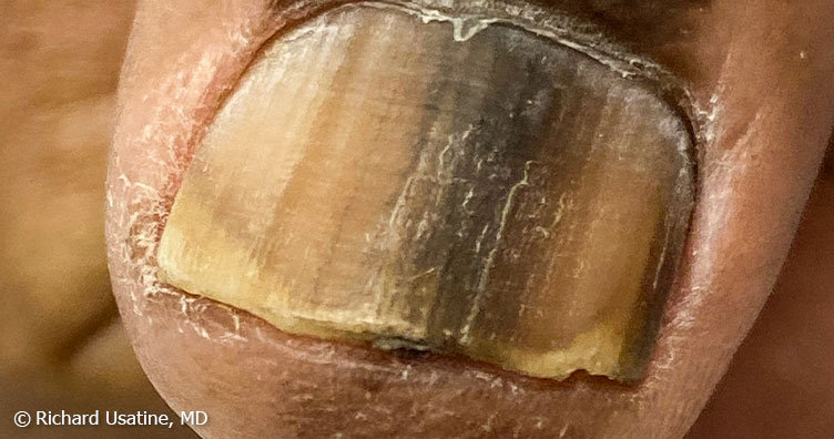
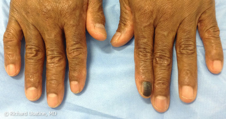
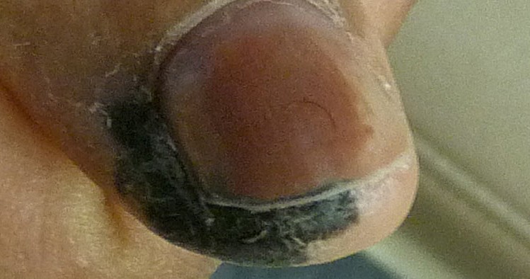
- Acral lentiginous melanoma (ALM)
- Cutaneous lymphoma
- Dermatofibrosarcoma protuberans (DFSP)
- Kaposi’s sarcoma
- Microcystic adnexal carcinoma (MAC)
- Sebaceous carcinoma
- Undifferentiated pleomorphic sarcoma
- Extramammary Paget’s disease (EMPD)
For links to more comprehensive skin cancer information, visit our Skin Cancer 101 page.
