Skin Cancer Pictures
What Does Skin Cancer Look Like?
Skin cancer can happen to anyone, at any age, on any part of the body. And because skin cancers appear in many shapes and sizes, they can be challenging to identify. While skin cancer pictures can be helpful in learning what skin cancer can look like, getting to know your own skin and understanding what to look for can help you detect cancer early when it’s easiest to cure.
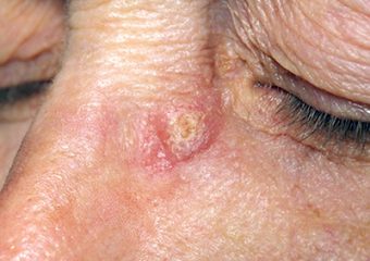
- Basal cell carcinoma (BCC) photos: Early and advanced BCCs, and BCCs on various skin tones.
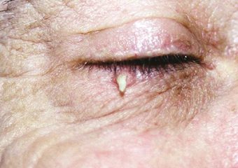
- Squamous cell carcinoma (SCC) photos: Early and late stage SCCs, and SCCs on various skin tones.
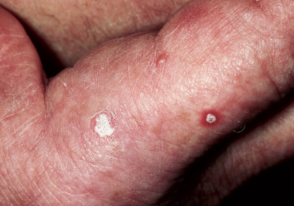
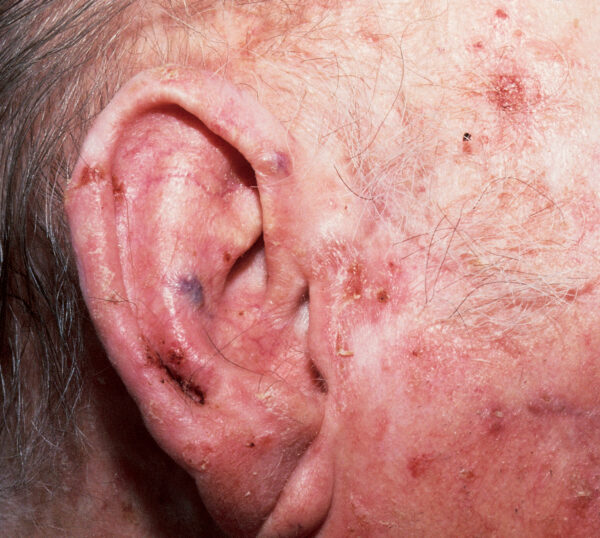
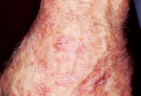
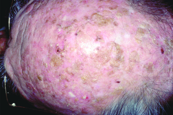
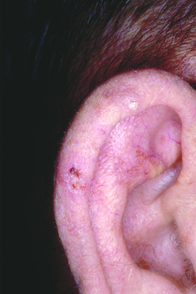
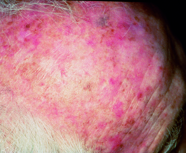
- Actinic keratosis (AK) photos: Images of this common precancer.
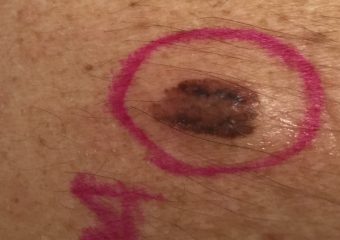
- Melanoma photos: Early stage and late stage melanoma images, and pictures of melanoma on various skin tones.
- Merkel cell carcinoma photos
To catch skin cancer early, you should examine your skin once a month. If you see something NEW, CHANGING OR UNUSUAL – even if it looks nothing like what you see in photos – do not wait! Get it checked by a dermatologist right away. Finding and treating skin cancer early can save your life.
Skin Cancer Image Gallery
What does cancer look like on skin? Below is a selection of photos that give you a general idea about what skin cancers can look like.
- Remember that skin cancers can look quite different from one person to another due to skin tone, size and type of skin cancer and location on the body.
- Skin cancer can be tricky in other ways, too. For example, melanoma is a type of skin cancer that is often pigmented tan, brown, black, even blue. But amelanotic melanoma lacks pigment and appears as a skin-tone or pink lesion.
To sum it up, while photos can be helpful, getting your skin examined by a dermatologist is the most vital step in identifying and treating skin cancer.
Please note: Since not all skin cancers have the same appearance, these photos serve as a general reference for what skin cancer can look like. If you see anything NEW, CHANGING or UNUSUAL on your skin, go get checked by a dermatologist.


[nested_image_filter]
Rare Skin Cancers
Please visit our Rare skin cancers page for more information and pictures of rare skin cancers such as:
- Acral lentiginous melanoma (ALM)
- Cutaneous lymphoma
- Dermatofibrosarcoma protuberans (DFSP)
- Kaposi’s sarcoma
- Microcystic adnexal carcinoma (MAC)
- Sebaceous carcinoma
- Undifferentiated pleomorphic sarcoma
- Extramammary Paget’s disease (EMPD)
For links to more comprehensive skin cancer information, visit our Skin Cancer 101 page.
